2C2I

Structure and function of Rv0130, a conserved hypothetical protein from M.tuberculosis


Experimental Data Snapshot

  • Method: X-RAY DIFFRACTION
  • Resolution: 1.80 Å
  • R-Value Free: 0.226 
  • R-Value Work: 0.183 
  • R-Value Observed: 0.185 

wwPDB Validation   3D Report Full Report


This is version 1.2 of the entry. See complete history


Literature

Structure and Function of Rv0130, a Conserved Hypothetical Protein from Mycobacterium Tuberculosis.

Johansson, P.Castell, A.Jones, T.A.Backbro, K.

(2006) Protein Sci 15: 2300

  • DOI: https://doi.org/10.1110/ps.062309306
  • Primary Citation of Related Structures:  
    2C2I

  • PubMed Abstract: 

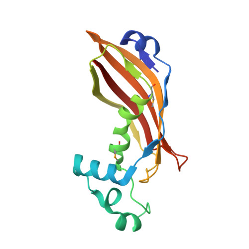
    A large fraction of the Mycobacterium tuberculosis genome codes for proteins of unknown function. We here report the structure of one of these proteins, Rv0130, solved to a resolution of 1.8 å. The Rv0130 monomer features a single hotdog fold composed of a highly curved beta-sheet on top of a long and a short alpha-helix. Two monomers in turn pack to form a double-hotdog-folded homodimer, similar to a large group of enzymes that use thiol esters as substrates. Rv0130 was found to contain a highly conserved R-specific hydratase motif buried deeply between the two monomers. Our biochemical studies show that the protein is able to hydrate a short trans-2-enoyl-coenzyme A moiety with a k(cat) of 1.1 x 10(2) sec(-1). The importance of the side chains of D40 and H45 for hydratase activity is demonstrated by site-directed mutagenesis. In contrast to many hotdog-folded proteins, a proline residue distorts the central helix of Rv0130. This distortion allows the creation of a long, curved tunnel, similar to the substrate-binding channels of long-chain eukaryotic hydratase 2 enzymes.


  • Organizational Affiliation

    Department of Cell and Molecular Biology, Uppsala University, Biomedical Center, SE-751 24 Uppsala, Sweden.


Macromolecules
Find similar proteins by:  (by identity cutoff)  |  3D Structure
Entity ID: 1
MoleculeChains Sequence LengthOrganismDetailsImage
RV0130
A, B
151Mycobacterium tuberculosis H37RvMutation(s): 0 
EC: 4.2.1.17
UniProt
Find proteins for P9WNP3 (Mycobacterium tuberculosis (strain ATCC 25618 / H37Rv))
Explore P9WNP3 
Go to UniProtKB:  P9WNP3
Entity Groups  
Sequence Clusters30% Identity50% Identity70% Identity90% Identity95% Identity100% Identity
UniProt GroupP9WNP3
Sequence Annotations
Expand
  • Reference Sequence
Small Molecules
Modified Residues  1 Unique
IDChains TypeFormula2D DiagramParent
MSE
Query on MSE
A, B
L-PEPTIDE LINKINGC5 H11 N O2 SeMET
Experimental Data & Validation

Experimental Data

  • Method: X-RAY DIFFRACTION
  • Resolution: 1.80 Å
  • R-Value Free: 0.226 
  • R-Value Work: 0.183 
  • R-Value Observed: 0.185 
  • Space Group: P 21 21 2
Unit Cell:
Length ( Å )Angle ( ˚ )
a = 58.697α = 90
b = 108.708β = 90
c = 50.376γ = 90
Software Package:
Software NamePurpose
REFMACrefinement

Structure Validation

View Full Validation Report


Entry History 

Deposition Data

Revision History  (Full details and data files)

  • Version 1.0: 2006-09-14
    Type: Initial release
  • Version 1.1: 2011-05-07
    Changes: Version format compliance
  • Version 1.2: 2011-07-13
    Changes: Version format compliance