Structural and Biochemical Characterisation of a Mitochondrial Peroxiredoxin from Plasmodium Falciparum
Boucher, I.W., Mcmillan, P.J., Gabrielsen, M., Akerman, S.E., Brannigan, J.A., Schnick, C., Brzozowski, A.M., Wilkinson, A.J., Muller, S.(2006) Mol Microbiol 61: 948
- PubMed: 16879648
- DOI: https://doi.org/10.1111/j.1365-2958.2006.05303.x
- Primary Citation of Related Structures:
2C0D - PubMed Abstract:
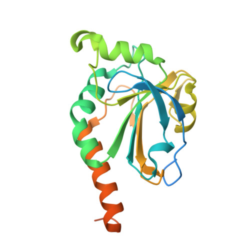
Plasmodium falciparum possesses a single mitochondrion with a functional electron transport chain. During respiration, reactive oxygen species are generated that need to be removed to protect the organelle from oxidative damage. In the absence of catalase and glutathione peroxidase, the parasites rely primarily on peroxiredoxin-linked systems for protection. We have analysed the biochemical and structural features of the mitochondrial peroxiredoxin and thioredoxin of P. falciparum. The mitochondrial localization of both proteins was confirmed by expressing green fluorescent protein fusions in parasite erythrocytic stages. Recombinant protein was kinetically characterized using the cytosolic and the mitochondrial thioredoxin (PfTrx1 and PfTrx2 respectively). The peroxiredoxin clearly preferred PfTrx2 to PfTrx1 as a reducing partner, reflected by the KM values of 11.6 microM and 130.4 microM respectively. Substitution of the two dyads asparagine-62/tyrosine-63 and phenylalanine-139/alanine-140 residues by aspartate-phenylalaine and valine-serine, respectively, reduced the KM for Trx1 but had no effect on the KM of Trx2 suggesting some role for these residues in the discrimination between the two substrates. Solution studies suggest that the protein exists primarily in a homodecameric form. The crystal structure of the mitochondrial peroxiredoxin reveals a fold typical of the 2-Cys class peroxiredoxins and a dimeric form with an intermolecular disulphide bridge between Cys67 and Cys187. These results show that the mitochondrial peroxiredoxin of P. falciparum occurs in both dimeric and decameric forms when purified under non-reducing conditions.
Organizational Affiliation:
Structural Biology Laboratory, Department of Chemistry, University of York, York YO10 5YW, UK.