Dodecameric Structure of the Small Heat Shock Protein Acr1 from Mycobacterium Tuberculosis.
Kennaway, C.K., Benesch, J.L.P., Gohlke, U., Wang, L., Robinson, C.V., Orlova, E.V., Saibil, H.R., Keep, N.H.(2005) J Biol Chem 280: 33419
- PubMed: 16046399
- DOI: https://doi.org/10.1074/jbc.M504263200
- Primary Citation of Related Structures:
2BYU - PubMed Abstract:
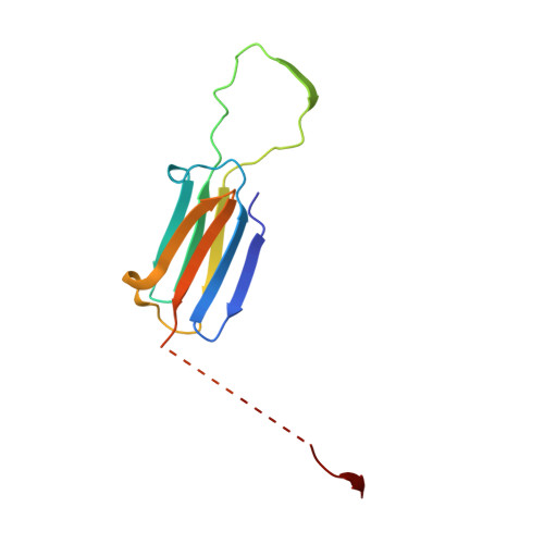
Small heat shock proteins are a ubiquitous and diverse family of stress proteins that have in common an alpha-crystallin domain. Mycobacterium tuberculosis has two small heat shock proteins, Acr1 (alpha-crystallin-related protein 1, or Hsp16.3/16-kDa antigen) and Acr2 (HrpA), both of which are highly expressed under different stress conditions. Small heat shock proteins form large oligomeric assemblies and are commonly polydisperse. Nanoelectrospray mass spectrometry showed that Acr2 formed a range of oligomers composed of dimers and tetramers, whereas Acr1 was a dodecamer. Electron microscopy of Acr2 showed a variety of particle sizes. Using three-dimensional analysis of negative stain electron microscope images, we have shown that Acr1 forms a tetrahedral assembly with 12 polypeptide chains. The atomic structure of a related alpha-crystallin domain dimer was docked into the density to build a molecular structure of the dodecameric Acr1 complex. Along with the differential regulation of these two proteins, the differences in their quaternary structures demonstrated here supports their distinct functional roles.
Organizational Affiliation:
School of Crystallography and Institute of Structural Molecular Biology, Birkbeck, University of London, Malet Street, London WC1E 7HX.