2BX9

Crystal structure of B.subtilis Anti-TRAP protein, an antagonist of TRAP-RNA interactions


Experimental Data Snapshot

  • Method: X-RAY DIFFRACTION
  • Resolution: 2.80 Å
  • R-Value Free: 0.296 
  • R-Value Work: 0.192 
  • R-Value Observed: 0.197 

wwPDB Validation   3D Report Full Report


This is version 1.1 of the entry. See complete history


Literature

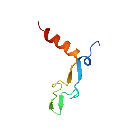
Crystal Structure of Bacillus Subtilis Anti-Trap Protein, an Antagonist of Trap/RNA Interaction.

Shevtsov, M.B.Chen, Y.Gollnick, P.Antson, A.A.

(2005) Proc Natl Acad Sci U S A 102: 17600

  • DOI: https://doi.org/10.1073/pnas.0508728102
  • Primary Citation of Related Structures:  
    2BX9

  • PubMed Abstract: 

    In Bacillus subtilis the anti-TRAP protein (AT) is produced in response to the accumulation of uncharged tRNA(Trp). AT regulates expression of genes involved in tryptophan biosynthesis and transport by binding to the tryptophan-activated trp RNA-binding attenuation protein (TRAP) and preventing its interaction with several mRNAs. Here, we report the x-ray structure of AT at 2.8 angstroms resolution, showing that the protein subunits assemble into tight trimers. Four such trimers are further associated into a 12-subunit particle in which individual trimers are related by twofold and threefold symmetry axes. Twelve DnaJ-like, cysteine-rich zinc-binding domains form spikes on the surface of the dodecamer. Available data suggest several possible ways for AT to interact with the 11-subunit TRAP. Interaction between the two symmetry-mismatching molecules could be assisted by the flexible nature of AT zinc-binding domains.


  • Organizational Affiliation

    York Structural Biology Laboratory, Department of Chemistry, University of York, York YO10 5YW, United Kingdom.


Macromolecules
Find similar proteins by:  (by identity cutoff)  |  3D Structure
Entity ID: 1
MoleculeChains Sequence LengthOrganismDetailsImage
TRYPTOPHAN RNA-BINDING ATTENUATOR PROTEIN-INHIBITORY PROTEIN
A, B, C, D, E
A, B, C, D, E, F, G, H, I, J, K, L
53Bacillus subtilisMutation(s): 0 
UniProt
Find proteins for O31466 (Bacillus subtilis (strain 168))
Explore O31466 
Go to UniProtKB:  O31466
Entity Groups  
Sequence Clusters30% Identity50% Identity70% Identity90% Identity95% Identity100% Identity
UniProt GroupO31466
Sequence Annotations
Expand
  • Reference Sequence
Small Molecules
Ligands 1 Unique
IDChains Name / Formula / InChI Key2D Diagram3D Interactions
ZN
Query on ZN

Download Ideal Coordinates CCD File 
M [auth A]
N [auth B]
O [auth C]
P [auth D]
Q [auth E]
M [auth A],
N [auth B],
O [auth C],
P [auth D],
Q [auth E],
R [auth F],
S [auth G],
T [auth H],
U [auth I],
V [auth J],
W [auth K],
X [auth L]
ZINC ION
Zn
PTFCDOFLOPIGGS-UHFFFAOYSA-N
Experimental Data & Validation

Experimental Data

  • Method: X-RAY DIFFRACTION
  • Resolution: 2.80 Å
  • R-Value Free: 0.296 
  • R-Value Work: 0.192 
  • R-Value Observed: 0.197 
  • Space Group: P 1
Unit Cell:
Length ( Å )Angle ( ˚ )
a = 51.552α = 113.99
b = 60.125β = 101.37
c = 60.348γ = 100.5
Software Package:
Software NamePurpose
MOSFLMdata reduction
SCALAdata scaling
SHELXDphasing
SHARPphasing
REFMACrefinement

Structure Validation

View Full Validation Report


Entry History 

Deposition Data

Revision History  (Full details and data files)

  • Version 1.0: 2005-11-30
    Type: Initial release
  • Version 1.1: 2011-07-13
    Changes: Advisory, Version format compliance