2BVN

E. coli EF-Tu:GDPNP in complex with the antibiotic enacyloxin IIa


Experimental Data Snapshot

  • Method: X-RAY DIFFRACTION
  • Resolution: 2.30 Å
  • R-Value Free: 0.271 
  • R-Value Work: 0.230 
  • R-Value Observed: 0.230 

wwPDB Validation   3D Report Full Report


This is version 1.6 of the entry. See complete history


Literature

Enacyloxin Iia Pinpoints a Binding Pocket of Elongation Factor TU for Development of Novel Antibiotics.

Parmeggiani, A.Krab, I.M.Watanabe, T.Nielsen, R.C.Dahlberg, C.Nyborg, J.Nissen, P.

(2006) J Biol Chem 281: 2893

  • DOI: https://doi.org/10.1074/jbc.M505951200
  • Primary Citation of Related Structures:  
    1OB5, 2BVN

  • PubMed Abstract: 

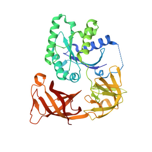
    Elongation factor (EF-) Tu.GTP is the carrier of aminoacyl-tRNA to the programmed ribosome. Enacyloxin IIa inhibits bacterial protein synthesis by hindering the release of EF-Tu.GDP from the ribosome. The crystal structure of the Escherichia coli EF-Tu.guanylyl iminodiphosphate (GDPNP).enacyloxin IIa complex at 2.3 A resolution presented here reveals the location of the antibiotic at the interface of domains 1 and 3. The binding site overlaps that of kirromycin, an antibiotic with a structure that is unrelated to enacyloxin IIa but that also inhibits EF-Tu.GDP release. As one of the major differences, the enacyloxin IIa tail borders a hydrophobic pocket that is occupied by the longer tail of kirromycin, explaining the higher binding affinity of the latter. EF-Tu.GDPNP.enacyloxin IIa shows a disordered effector region that in the Phe-tRNAPhe.EF-Tu (Thermus aquaticus).GDPNP.enacyloxin IIa complex, solved at 3.1 A resolution, is stabilized by the interaction with tRNA. This work clarifies the structural background of the action of enacyloxin IIa and compares its properties with those of kirromycin, opening new perspectives for structure-guided design of novel antibiotics.


  • Organizational Affiliation

    Department of Molecular Biology, University of Aarhus, Gustav Wieds Vej 10 C, DK-8000 Aarhus C, Denmark. andrea@bioxray.dk


Macromolecules
Find similar proteins by:  (by identity cutoff)  |  3D Structure
Entity ID: 1
MoleculeChains Sequence LengthOrganismDetailsImage
ELONGATION FACTOR TU
A, B
393Escherichia coli BL21Mutation(s): 0 
EC: 3.6.1.48
UniProt
Find proteins for P0CE48 (Escherichia coli (strain K12))
Explore P0CE48 
Go to UniProtKB:  P0CE48
Entity Groups  
Sequence Clusters30% Identity50% Identity70% Identity90% Identity95% Identity100% Identity
UniProt GroupP0CE48
Sequence Annotations
Expand
  • Reference Sequence
Experimental Data & Validation

Experimental Data

  • Method: X-RAY DIFFRACTION
  • Resolution: 2.30 Å
  • R-Value Free: 0.271 
  • R-Value Work: 0.230 
  • R-Value Observed: 0.230 
  • Space Group: P 43 21 2
Unit Cell:
Length ( Å )Angle ( ˚ )
a = 72.07α = 90
b = 72.07β = 90
c = 335.73γ = 90
Software Package:
Software NamePurpose
CNSrefinement
DENZOdata reduction
SCALEPACKdata scaling
AMoREphasing

Structure Validation

View Full Validation Report


Entry History 

Deposition Data

Revision History  (Full details and data files)

  • Version 1.0: 2005-09-01
    Type: Initial release
  • Version 1.1: 2011-05-08
    Changes: Version format compliance
  • Version 1.2: 2011-07-13
    Changes: Version format compliance
  • Version 1.3: 2018-01-17
    Changes: Data collection
  • Version 1.4: 2019-05-08
    Changes: Data collection, Experimental preparation
  • Version 1.5: 2019-05-15
    Changes: Data collection, Experimental preparation
  • Version 1.6: 2023-12-13
    Changes: Data collection, Database references, Derived calculations, Refinement description