2BVC

Crystal structure of Mycobacterium tuberculosis glutamine synthetase in complex with a transition state mimic


Experimental Data Snapshot

  • Method: X-RAY DIFFRACTION
  • Resolution: 2.10 Å
  • R-Value Free: 0.230 
  • R-Value Work: 0.191 
  • R-Value Observed: 0.193 

wwPDB Validation   3D Report Full Report


Ligand Structure Quality Assessment 


This is version 1.3 of the entry. See complete history


Literature

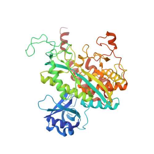
Structure of Mycobacterium Tuberculosis Glutamine Synthetase in Complex with a Transition-State Mimic Provides Functional Insights.

Krajewski, W.W.Jones, T.A.Mowbray, S.L.

(2005) Proc Natl Acad Sci U S A 102: 10499

  • DOI: https://doi.org/10.1073/pnas.0502248102
  • Primary Citation of Related Structures:  
    2BVC

  • PubMed Abstract: 

    Glutamine synthetase catalyzes the ligation of glutamate and ammonia to form glutamine, with the resulting hydrolysis of ATP. The enzyme is a central component of bacterial nitrogen metabolism and is a potential drug target. Here, we report a high-yield recombinant expression system for glutamine synthetase of Mycobacterium tuberculosis together with a simple purification. The procedure allowed the structure of a complex with a phosphorylated form of the inhibitor methionine sulfoximine, magnesium, and ADP to be solved by molecular replacement and refined at 2.1-A resolution. To our knowledge, this study provides the first reported structure for a taut form of the M. tuberculosis enzyme, similar to that observed for the Salmonella enzyme earlier. The phospho compound, generated in situ by an active enzyme, mimics the phosphorylated tetrahedral adduct at the transition state. Some differences in ligand interactions of the protein with both phosphorylated compound and nucleotide are observed compared with earlier structures; a third metal ion also is found. The importance of these differences in the catalytic mechanism is discussed; the results will help guide the search for specific inhibitors of potential therapeutic interest.


  • Organizational Affiliation

    Department of Cell and Molecular Biology, Uppsala University, Biomedical Center, Box 596, SE-751 24 Uppsala, Sweden.


Macromolecules
Find similar proteins by:  (by identity cutoff)  |  3D Structure
Entity ID: 1
MoleculeChains Sequence LengthOrganismDetailsImage
GLUTAMINE SYNTHETASE 1
A, B, C, D, E
A, B, C, D, E, F
486Mycobacterium tuberculosis H37RvMutation(s): 0 
EC: 6.3.1.2
UniProt
Find proteins for P9WN39 (Mycobacterium tuberculosis (strain ATCC 25618 / H37Rv))
Explore P9WN39 
Go to UniProtKB:  P9WN39
Entity Groups  
Sequence Clusters30% Identity50% Identity70% Identity90% Identity95% Identity100% Identity
UniProt GroupP9WN39
Sequence Annotations
Expand
  • Reference Sequence
Small Molecules
Ligands 4 Unique
IDChains Name / Formula / InChI Key2D Diagram3D Interactions
ADP
Query on ADP

Download Ideal Coordinates CCD File 
FA [auth E]
H [auth A]
LA [auth F]
N [auth B]
T [auth C]
FA [auth E],
H [auth A],
LA [auth F],
N [auth B],
T [auth C],
Z [auth D]
ADENOSINE-5'-DIPHOSPHATE
C10 H15 N5 O10 P2
XTWYTFMLZFPYCI-KQYNXXCUSA-N
P3S
Query on P3S

Download Ideal Coordinates CCD File 
EA [auth E]
G [auth A]
KA [auth F]
M [auth B]
S [auth C]
EA [auth E],
G [auth A],
KA [auth F],
M [auth B],
S [auth C],
Y [auth D]
L-METHIONINE-S-SULFOXIMINE PHOSPHATE
C5 H13 N2 O6 P S
QQFOFBSCSWFFPB-NMAPHRJESA-N
CL
Query on CL

Download Ideal Coordinates CCD File 
DA [auth D]
JA [auth E]
L [auth A]
PA [auth F]
R [auth B]
DA [auth D],
JA [auth E],
L [auth A],
PA [auth F],
R [auth B],
X [auth C]
CHLORIDE ION
Cl
VEXZGXHMUGYJMC-UHFFFAOYSA-M
MG
Query on MG

Download Ideal Coordinates CCD File 
AA [auth D]
BA [auth D]
CA [auth D]
GA [auth E]
HA [auth E]
AA [auth D],
BA [auth D],
CA [auth D],
GA [auth E],
HA [auth E],
I [auth A],
IA [auth E],
J [auth A],
K [auth A],
MA [auth F],
NA [auth F],
O [auth B],
OA [auth F],
P [auth B],
Q [auth B],
U [auth C],
V [auth C],
W [auth C]
MAGNESIUM ION
Mg
JLVVSXFLKOJNIY-UHFFFAOYSA-N
Experimental Data & Validation

Experimental Data

  • Method: X-RAY DIFFRACTION
  • Resolution: 2.10 Å
  • R-Value Free: 0.230 
  • R-Value Work: 0.191 
  • R-Value Observed: 0.193 
  • Space Group: C 2 2 21
Unit Cell:
Length ( Å )Angle ( ˚ )
a = 133.261α = 90
b = 229.457β = 90
c = 203.167γ = 90
Software Package:
Software NamePurpose
REFMACrefinement
DENZOdata reduction
SCALEPACKdata scaling
MOLREPphasing

Structure Validation

View Full Validation Report


Ligand Structure Quality Assessment 


Entry History 

Deposition Data

Revision History  (Full details and data files)

  • Version 1.0: 2005-07-07
    Type: Initial release
  • Version 1.1: 2011-05-08
    Changes: Version format compliance
  • Version 1.2: 2011-07-13
    Changes: Version format compliance
  • Version 1.3: 2023-12-13
    Changes: Data collection, Database references, Derived calculations, Other, Refinement description