2BUV

Crystal Structure Of Protocatechuate 3,4-Dioxygenase from Acinetobacter Sp. ADP1 Mutant R457S in Complex with Protocatechuate


Experimental Data Snapshot

  • Method: X-RAY DIFFRACTION
  • Resolution: 1.80 Å
  • R-Value Free: 0.202 
  • R-Value Work: 0.174 
  • R-Value Observed: 0.174 

wwPDB Validation   3D Report Full Report


Ligand Structure Quality Assessment 


This is version 1.5 of the entry. See complete history


Literature

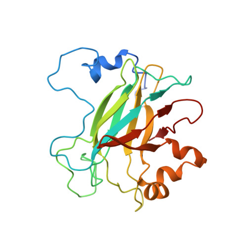
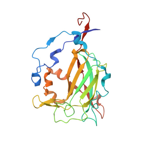
Biophysical Analyses of Designed and Selected Mutants of Protocatechuate 3,4-Dioxygenase

Brown, C.K.Vetting, M.W.Earhart, C.A.Ohlendorf, D.H.

(2004) Annu Rev Microbiol 58: 555-585

  • DOI: https://doi.org/10.1146/annurev.micro.57.030502.090927
  • Primary Citation of Related Structures:  
    2BUM, 2BUQ, 2BUR, 2BUT, 2BUV

  • PubMed Abstract: 

    The catechol dioxygenases allow a wide variety of bacteria to use aromatic compounds as carbon sources by catalyzing the key ring-opening step. These enzymes use specifically either catechol or protocatechuate (2,3-dihydroxybenozate) as their substrates; they use a bare metal ion as the sole cofactor. To learn how this family of metalloenzymes functions, a structural analysis of designed and selected mutants of these enzymes has been undertaken. Here we review the results of this analysis on the nonheme ferric iron intradiol dioxygenase protocatechuate 3,4-dioxygenase.


  • Organizational Affiliation

    Center for Metals in Biocatalysis and Department of Biochemistry, Molecular Biology, and Biophysics , Minneapolis, Minnesota 55455, USA. brow0927@umn.edu


Macromolecules
Find similar proteins by:  (by identity cutoff)  |  3D Structure
Entity ID: 1
MoleculeChains Sequence LengthOrganismDetailsImage
PROTOCATECHUATE 3,4-DIOXYGENASE ALPHA CHAIN209Acinetobacter baylyi ADP1Mutation(s): 0 
EC: 1.13.11.3
UniProt
Find proteins for P20371 (Acinetobacter baylyi (strain ATCC 33305 / BD413 / ADP1))
Explore P20371 
Go to UniProtKB:  P20371
Entity Groups  
Sequence Clusters30% Identity50% Identity70% Identity90% Identity95% Identity100% Identity
UniProt GroupP20371
Sequence Annotations
Expand
  • Reference Sequence
Find similar proteins by:  (by identity cutoff)  |  3D Structure
Entity ID: 2
MoleculeChains Sequence LengthOrganismDetailsImage
PROTOCATECHUATE 3,4-DIOXYGENASE BETA CHAIN241Acinetobacter baylyi ADP1Mutation(s): 1 
EC: 1.13.11.3
UniProt
Find proteins for P20372 (Acinetobacter baylyi (strain ATCC 33305 / BD413 / ADP1))
Explore P20372 
Go to UniProtKB:  P20372
Entity Groups  
Sequence Clusters30% Identity50% Identity70% Identity90% Identity95% Identity100% Identity
UniProt GroupP20372
Sequence Annotations
Expand
  • Reference Sequence
Small Molecules
Binding Affinity Annotations 
IDSourceBinding Affinity
DHB Binding MOAD:  2BUV Kd: 1.00e+5 (nM) from 1 assay(s)
Experimental Data & Validation

Experimental Data

  • Method: X-RAY DIFFRACTION
  • Resolution: 1.80 Å
  • R-Value Free: 0.202 
  • R-Value Work: 0.174 
  • R-Value Observed: 0.174 
  • Space Group: I 2 3
Unit Cell:
Length ( Å )Angle ( ˚ )
a = 144.7α = 90
b = 144.7β = 90
c = 144.7γ = 90
Software Package:
Software NamePurpose
CNSrefinement
CrystalCleardata reduction
CrystalCleardata scaling
CNSphasing

Structure Validation

View Full Validation Report


Ligand Structure Quality Assessment 


Entry History 

Deposition Data

Revision History  (Full details and data files)

  • Version 1.0: 2006-09-04
    Type: Initial release
  • Version 1.1: 2011-05-08
    Changes: Version format compliance
  • Version 1.2: 2011-07-13
    Changes: Version format compliance
  • Version 1.3: 2018-06-20
    Changes: Data collection, Database references
  • Version 1.4: 2019-05-22
    Changes: Data collection, Refinement description
  • Version 1.5: 2023-12-13
    Changes: Data collection, Database references, Derived calculations, Other, Refinement description