2BUB

Crystal Structure Of Human Dipeptidyl Peptidase IV (CD26) in Complex with a Reversed Amide Inhibitor


Experimental Data Snapshot

  • Method: X-RAY DIFFRACTION
  • Resolution: 2.66 Å
  • R-Value Free: 0.329 
  • R-Value Work: 0.258 
  • R-Value Observed: 0.260 

wwPDB Validation   3D Report Full Report


This is version 2.4 of the entry. See complete history


Literature

The reversed binding of beta-phenethylamine inhibitors of DPP-IV: X-ray structures and properties of novel fragment and elaborated inhibitors.

Nordhoff, S.Cerezo-Galvez, S.Feurer, A.Hill, O.Matassa, V.G.Metz, G.Rummey, C.Thiemann, M.Edwards, P.J.

(2006) Bioorg Med Chem Lett 16: 1744-1748

  • DOI: https://doi.org/10.1016/j.bmcl.2005.11.103
  • Primary Citation of Related Structures:  
    2BUA, 2BUB, 2BUC

  • PubMed Abstract: 

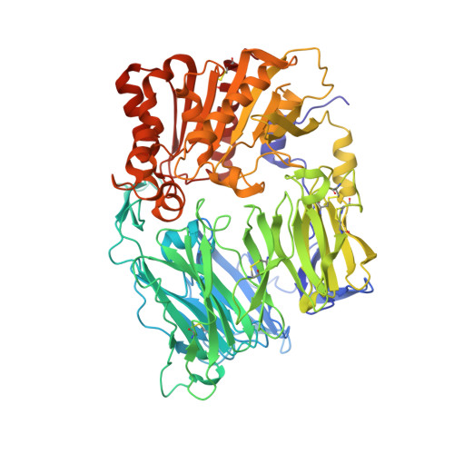
    The co-crystal structure of beta-phenethylamine fragment inhibitor 5 bound to DPP-IV revealed that the phenyl ring occupied the proline pocket of the enzyme. This finding provided the basis for a general hypothesis of a reverse binding mode for beta-phenethylamine-based DPP-IV inhibitors. Novel inhibitor design concepts that obviate substrate-like structure-activity relationships (SAR) were thereby enabled, and novel, potent inhibitors were discovered.


  • Organizational Affiliation

    Medicinal Chemistry, Santhera Pharmaceuticals, Im Neuenheimer Feld 518-519, D-69120 Heidelberg, Germany. sonja.nordhoff@santhera.com


Macromolecules
Find similar proteins by:  (by identity cutoff)  |  3D Structure
Entity ID: 1
MoleculeChains Sequence LengthOrganismDetailsImage
DIPEPTIDYL PEPTIDASE 4
A, B
728Homo sapiensMutation(s): 0 
EC: 3.4.14.5
UniProt & NIH Common Fund Data Resources
Find proteins for P27487 (Homo sapiens)
Explore P27487 
Go to UniProtKB:  P27487
PHAROS:  P27487
GTEx:  ENSG00000197635 
Entity Groups  
Sequence Clusters30% Identity50% Identity70% Identity90% Identity95% Identity100% Identity
UniProt GroupP27487
Sequence Annotations
Expand
  • Reference Sequence
Small Molecules
Binding Affinity Annotations 
IDSourceBinding Affinity
FPB BindingDB:  2BUB IC50: min: 90, max: 410 (nM) from 2 assay(s)
PDBBind:  2BUB IC50: 90 (nM) from 1 assay(s)
Experimental Data & Validation

Experimental Data

  • Method: X-RAY DIFFRACTION
  • Resolution: 2.66 Å
  • R-Value Free: 0.329 
  • R-Value Work: 0.258 
  • R-Value Observed: 0.260 
  • Space Group: P 21 21 21
Unit Cell:
Length ( Å )Angle ( ˚ )
a = 65.486α = 90
b = 66.773β = 90
c = 425.407γ = 90
Software Package:
Software NamePurpose
REFMACrefinement

Structure Validation

View Full Validation Report


Entry History 

Deposition Data

Revision History  (Full details and data files)

  • Version 1.0: 2006-01-23
    Type: Initial release
  • Version 1.1: 2011-05-08
    Changes: Version format compliance
  • Version 1.2: 2011-07-13
    Changes: Version format compliance
  • Version 2.0: 2018-08-22
    Changes: Advisory, Atomic model, Data collection, Database references, Derived calculations, Non-polymer description, Structure summary
  • Version 2.1: 2019-07-10
    Changes: Data collection, Derived calculations
  • Version 2.2: 2019-07-24
    Changes: Data collection
  • Version 2.3: 2019-09-04
    Changes: Data collection
  • Version 2.4: 2020-07-29
    Type: Remediation
    Reason: Carbohydrate remediation
    Changes: Data collection, Derived calculations, Structure summary