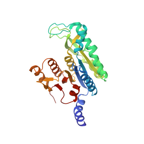
Structural Bases of Feed-Back Control of Arginine Biosynthesis, Revealed by the Structure of Two Hexameric N-Acetylglutamate Kinases, from Thermotoga Maritima and Pseudomonas Aeruginosa
Ramon-Maiques, S., Fernandez-Murga, M.L., Gil-Ortiz, F., Vagin, A., Fita, I., Rubio, V.(2006) J Mol Biol 356: 695
- PubMed: 16376937
- DOI: https://doi.org/10.1016/j.jmb.2005.11.079
- Primary Citation of Related Structures:
2BTY, 2BUF - PubMed Abstract:
N-Acetylglutamate kinase (NAGK) catalyses the second step in the route of arginine biosynthesis. In many organisms this enzyme is inhibited by the final product of the route, arginine, and thus plays a central regulatory role. In addition, in photosynthetic organisms NAGK is the target of the nitrogen-signalling protein PII. The 3-D structure of homodimeric, arginine-insensitive, Escherichia coli NAGK, clarified substrate binding and catalysis but shed no light on arginine inhibition of NAGK. We now shed light on arginine inhibition by determining the crystal structures, at 2.75 A and 2.95 A resolution, of arginine-complexed Thermotoga maritima and arginine-free Pseudomonas aeruginosa NAGKs, respectively. Both enzymes are highly similar ring-like hexamers having a central orifice of approximately 30 A diameter. They are formed by linking three E.coli NAGK-like homodimers through the interlacing of an N-terminal mobile kinked alpha-helix, which is absent from E.coli NAGK. Arginine is bound in each subunit of T.maritima NAGK, flanking the interdimeric junction, in a site formed between the N helix and the C lobe of the subunit. This site is also present, in variable conformations, in P.aeruginosa NAGK, but is missing from E.coli NAGK. Arginine, by gluing the C lobe of each subunit to the inter-dimeric junction, may stabilize an enlarged active centre conformation, hampering catalysis. Acetylglutamate counters arginine inhibition by promoting active centre closure. The hexameric architecture justifies the observed sigmoidal arginine inhibition kinetics with a high Hill coefficient (N approximately 4), and appears essential for arginine inhibition and for NAGK-PII complex formation, since this complex may involve binding of NAGK and PII with their 3-fold axes aligned. The NAGK structures allow identification of diagnostic sequence signatures for arginine inhibition. These signatures are found also in the homologous arginine-inhibited enzyme NAG synthase. The findings on NAGK shed light on the structure, function and arginine inhibition of this synthase, for which a hexameric model is constructed.
Organizational Affiliation:
Instituto de Biomedicina de Valencia (CSIC), Jaume Roig 11, Valencia 46010, Spain.