


Crystal Structure of Phosphoribosylformylglycinamidine Cyclo-Ligase from Bacillus Anthracis at 2.3A Resolution.
Moroz, O.V., Blagova, E.V., Levdikov, V.M., Fogg, M.J., Lebedev, A.A., Brannigan, J.A., Wilkinson, A.J., Wilson, K.S.To be published.
Experimental Data Snapshot
wwPDB Validation 3D Report Full Report

Entity ID: 1 | |||||
|---|---|---|---|---|---|
| Molecule | Chains | Sequence Length | Organism | Details | Image |
| PHOSPHORIBOSYL-AMINOIMIDAZOLE SYNTHETASE | 346 | Bacillus anthracis | Mutation(s): 0 EC: 6.3.3.1 | 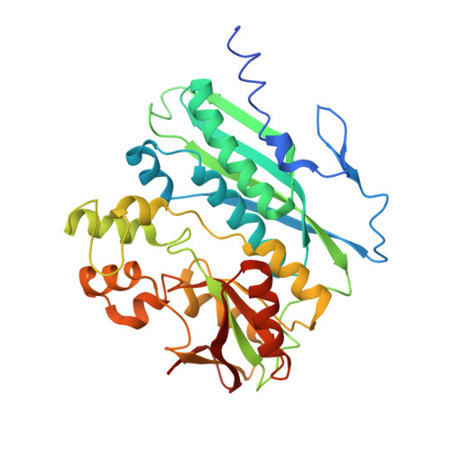 | |
UniProt | |||||
Find proteins for Q81ZH0 (Bacillus anthracis) Explore Q81ZH0 Go to UniProtKB: Q81ZH0 | |||||
Entity Groups | |||||
| Sequence Clusters | 30% Identity50% Identity70% Identity90% Identity95% Identity100% Identity | ||||
| UniProt Group | Q81ZH0 | ||||
Sequence AnnotationsExpand | |||||
| |||||
| Length ( Å ) | Angle ( ˚ ) |
|---|---|
| a = 89.49 | α = 90 |
| b = 89.49 | β = 90 |
| c = 88.117 | γ = 90 |
| Software Name | Purpose |
|---|---|
| SHELXL-97 | refinement |
| DENZO | data reduction |
| SCALEPACK | data scaling |
| MOLREP | phasing |
RCSB PDB (citation) is hosted by
RCSB PDB is a member of the

