2BSK

Crystal structure of the TIM9 Tim10 hexameric complex


Experimental Data Snapshot

  • Method: X-RAY DIFFRACTION
  • Resolution: 3.30 Å
  • R-Value Free: 0.319 
  • R-Value Work: 0.268 
  • R-Value Observed: 0.268 

wwPDB Validation   3D Report Full Report


This is version 1.2 of the entry. See complete history


Literature

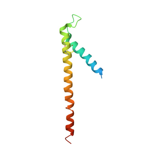
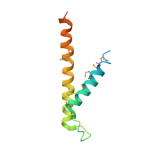
Crystal Structure of the Mitochondrial Chaperone Tim910 Reveals a Six-Bladed Alpha-Propeller.

Webb, C.T.Gorman, M.A.Lazarou, M.Ryan, M.T.Gulbis, J.M.

(2006) Mol Cell 21: 123

  • DOI: https://doi.org/10.1016/j.molcel.2005.11.010
  • Primary Citation of Related Structures:  
    2BSK

  • PubMed Abstract: 

    Import of proteins into mitochondria occurs by coordinated actions of preprotein translocases in the outer and inner membranes. Tim9 and Tim10 are translocase components of the intermembrane space, related to deafness-dystonia peptide 1 (DDP1). They coassemble into a hexamer, TIM9.10, which captures and chaperones precursors of inner membrane metabolite carriers as they exit the TOM channel in the outer membrane. The crystal structure of TIM9.10 reveals a previously undescribed alpha-propeller topology in which helical "blades" radiate from a narrow central pore lined with polar residues. The propeller blades are reminiscent of "tentacles" in chaperones Skp and prefoldin. In each TIM9.10 subunit, a signature "twin CX3C" motif forms two intramolecular disulfides. There is no obvious binding pocket for precursors, which we suggest employ the chaperone-like tentacles of TIM9.10 as surrogate lipid contacts. The first reported crystal structure of a mitochondrial translocase assembly provides insights into selectivity and regulation of precursor import.


  • Organizational Affiliation

    Structural Biology Division, The Walter and Eliza Hall Institute of Medical Research, 1G Royal Parade, Parkville, Victoria 3050, Australia.


Macromolecules
Find similar proteins by:  (by identity cutoff)  |  3D Structure
Entity ID: 1
MoleculeChains Sequence LengthOrganismDetailsImage
MITOCHONDRIAL IMPORT INNER MEMBRANE TRANSLOCASE SUBUNIT TIM9 A
A, C, E
89Homo sapiensMutation(s): 0 
UniProt & NIH Common Fund Data Resources
Find proteins for Q9Y5J7 (Homo sapiens)
Explore Q9Y5J7 
Go to UniProtKB:  Q9Y5J7
PHAROS:  Q9Y5J7
GTEx:  ENSG00000100575 
Entity Groups  
Sequence Clusters30% Identity50% Identity70% Identity90% Identity95% Identity100% Identity
UniProt GroupQ9Y5J7
Sequence Annotations
Expand
  • Reference Sequence
Find similar proteins by:  (by identity cutoff)  |  3D Structure
Entity ID: 2
MoleculeChains Sequence LengthOrganismDetailsImage
MITOCHONDRIAL IMPORT INNER MEMBRANE TRANSLOCASE SUBUNIT TIM10
B, D, F
90Homo sapiensMutation(s): 0 
UniProt & NIH Common Fund Data Resources
Find proteins for P62072 (Homo sapiens)
Explore P62072 
Go to UniProtKB:  P62072
PHAROS:  P62072
GTEx:  ENSG00000134809 
Entity Groups  
Sequence Clusters30% Identity50% Identity70% Identity90% Identity95% Identity100% Identity
UniProt GroupP62072
Sequence Annotations
Expand
  • Reference Sequence
Small Molecules
Modified Residues  1 Unique
IDChains TypeFormula2D DiagramParent
MSE
Query on MSE
B, D, F
L-PEPTIDE LINKINGC5 H11 N O2 SeMET
Experimental Data & Validation

Experimental Data

  • Method: X-RAY DIFFRACTION
  • Resolution: 3.30 Å
  • R-Value Free: 0.319 
  • R-Value Work: 0.268 
  • R-Value Observed: 0.268 
  • Space Group: P 41 21 2
Unit Cell:
Length ( Å )Angle ( ˚ )
a = 107.43α = 90
b = 107.43β = 90
c = 110.588γ = 90
Software Package:
Software NamePurpose
CNSrefinement
HKL-2000data reduction
HKL-2000data scaling
RESOLVEphasing

Structure Validation

View Full Validation Report


Entry History 

Deposition Data

Revision History  (Full details and data files)

  • Version 1.0: 2006-01-04
    Type: Initial release
  • Version 1.1: 2011-05-08
    Changes: Version format compliance
  • Version 1.2: 2011-07-13
    Changes: Version format compliance