2BSG

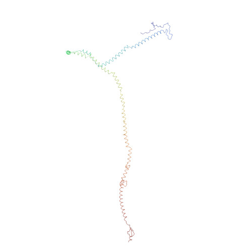
The modeled structure of fibritin (gpwac) of bacteriophage T4 based on cryo-EM reconstruction of the extended tail of bacteriophage T4


Experimental Data Snapshot

  • Method: ELECTRON MICROSCOPY
  • Resolution: 15.0 Å
  • Aggregation State: FILAMENT 
  • Reconstruction Method: HELICAL 

wwPDB Validation   3D Report Full Report


This is version 1.3 of the entry. See complete history


Literature

The Tail Structure of Bacteriophage T4 and its Mechanism of Contraction.

Kostyuchenko, V.A.Chipman, P.R.Leiman, P.G.Arisaka, F.Mesyanzhinov, V.V.Rossmann, M.G.

(2005) Nat Struct Mol Biol 12: 810

  • DOI: https://doi.org/10.1038/nsmb975
  • Primary Citation of Related Structures:  
    1ZKU, 2BSG

  • PubMed Abstract: 

    Bacteriophage T4 and related viruses have a contractile tail that serves as an efficient mechanical device for infecting bacteria. A three-dimensional cryo-EM reconstruction of the mature T4 tail assembly at 15-A resolution shows the hexagonal dome-shaped baseplate, the extended contractile sheath, the long tail fibers attached to the baseplate and the collar formed by six whiskers that interact with the long tail fibers. Comparison with the structure of the contracted tail shows that tail contraction is associated with a substantial rearrangement of the domains within the sheath protein and results in shortening of the sheath to about one-third of its original length. During contraction, the tail tube extends beneath the baseplate by about one-half of its total length and rotates by 345 degrees , allowing it to cross the host's periplasmic space.


  • Organizational Affiliation

    Department of Biological Sciences, Purdue University, 915 W. State Street, West Lafayette, Indiana 47907-2054, USA.


Macromolecules
Find similar proteins by:  (by identity cutoff)  |  3D Structure
Entity ID: 1
MoleculeChains Sequence LengthOrganismDetailsImage
FIBRITIN
A, B, C
487Tequatrovirus T4Mutation(s): 0 
UniProt
Find proteins for P10104 (Enterobacteria phage T4)
Explore P10104 
Go to UniProtKB:  P10104
Entity Groups  
Sequence Clusters30% Identity50% Identity70% Identity90% Identity95% Identity100% Identity
UniProt GroupP10104
Sequence Annotations
Expand
  • Reference Sequence
Experimental Data & Validation

Experimental Data

  • Method: ELECTRON MICROSCOPY
  • Resolution: 15.0 Å
  • Aggregation State: FILAMENT 
  • Reconstruction Method: HELICAL 
EM Software:
TaskSoftware PackageVersion
RECONSTRUCTIONSPIDER

Structure Validation

View Full Validation Report


Entry History 

Deposition Data

Revision History  (Full details and data files)

  • Version 1.0: 2005-09-21
    Type: Initial release
  • Version 1.1: 2014-01-15
    Changes: Database references, Derived calculations, Other, Version format compliance
  • Version 1.2: 2017-08-30
    Changes: Data collection, Experimental preparation
  • Version 1.3: 2019-10-23
    Changes: Data collection, Other