2BON

Structure of an Escherichia coli lipid kinase (YegS)


Experimental Data Snapshot

  • Method: X-RAY DIFFRACTION
  • Resolution: 1.90 Å
  • R-Value Free: 0.267 
  • R-Value Work: 0.220 
  • R-Value Observed: 0.222 

wwPDB Validation   3D Report Full Report


This is version 1.1 of the entry. See complete history


Literature

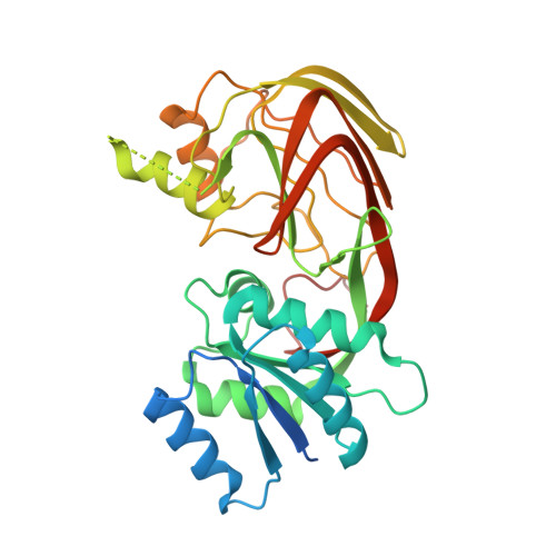
Crystal Structure of Yegs, a Homologue to the Mammalian Diacylglycerol Kinases, Reveals a Novel Regulatory Metal Binding Site.

Bakali, H.M.Herman, M.D.Johnson, K.A.Kelly, A.A.Wieslander, A.Hallberg, B.M.Nordlund, P.

(2007) J Biol Chem 282: 19644

  • DOI: https://doi.org/10.1074/jbc.M604852200
  • Primary Citation of Related Structures:  
    2BON, 2JGR

  • PubMed Abstract: 

    The human lipid kinase family controls cell proliferation, differentiation, and tumorigenesis and includes diacylglycerol kinases, sphingosine kinases, and ceramide kinases. YegS is an Escherichia coli protein with significant sequence homology to the catalytic domain of the human lipid kinases. We have solved the crystal structure of YegS and shown that it is a lipid kinase with phosphatidylglycerol kinase activity. The crystal structure reveals a two-domain protein with significant structural similarity to a family of NAD kinases. The active site is located in the interdomain cleft formed by four conserved sequence motifs. Surprisingly, the structure reveals a novel metal binding site composed of residues conserved in most lipid kinases.


  • Organizational Affiliation

    Division of Biophysics, Department of Medical Biochemistry and Biophysics, Karolinska Institute, SE-171 77 Stockholm, Sweden.


Macromolecules
Find similar proteins by:  (by identity cutoff)  |  3D Structure
Entity ID: 1
MoleculeChains Sequence LengthOrganismDetailsImage
LIPID KINASE
A, B
332Escherichia coli BL21(DE3)Mutation(s): 0 
UniProt
Find proteins for P76407 (Escherichia coli (strain K12))
Explore P76407 
Go to UniProtKB:  P76407
Entity Groups  
Sequence Clusters30% Identity50% Identity70% Identity90% Identity95% Identity100% Identity
UniProt GroupP76407
Sequence Annotations
Expand
  • Reference Sequence
Experimental Data & Validation

Experimental Data

  • Method: X-RAY DIFFRACTION
  • Resolution: 1.90 Å
  • R-Value Free: 0.267 
  • R-Value Work: 0.220 
  • R-Value Observed: 0.222 
  • Space Group: P 1 21 1
Unit Cell:
Length ( Å )Angle ( ˚ )
a = 42.326α = 90
b = 166.164β = 97.03
c = 48.474γ = 90
Software Package:
Software NamePurpose
REFMACrefinement
MOSFLMdata reduction
SCALAdata scaling
SHELXDphasing
SOLVEphasing
RESOLVEphasing

Structure Validation

View Full Validation Report


Entry History 

Deposition Data

Revision History  (Full details and data files)

  • Version 1.0: 2006-10-18
    Type: Initial release
  • Version 1.1: 2011-07-13
    Changes: Advisory, Version format compliance