2BOJ

crystal Structure of pseudomonas aeruginosa lectin (PA-IIL) complexed with methyl-B-D-Arabinopyranoside


Experimental Data Snapshot

  • Method: X-RAY DIFFRACTION
  • Resolution: 1.80 Å
  • R-Value Free: 0.172 
  • R-Value Work: 0.136 
  • R-Value Observed: 0.137 

wwPDB Validation   3D Report Full Report


Ligand Structure Quality Assessment 


This is version 1.4 of the entry. See complete history


Literature

Binding of Different Monosaccharides by Lectin Pa-Iil from Pseudoman Aeruginosa: Thermodynamics Data Correlated with X-Ray Structures.

Sabin, C.D.Mitchell, E.P.Pokarna, M.Gautier, C.Utille, J.-P.Wimmerova, M.Imberty, A.

(2006) FEBS Lett 580: 982

  • DOI: https://doi.org/10.1016/j.febslet.2006.01.030
  • Primary Citation of Related Structures:  
    2BOJ, 2BP6

  • PubMed Abstract: 

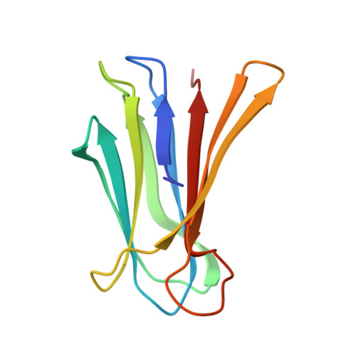
    The lectin from Pseudomonas aeruginosa (PA-IIL) is involved in host recognition and biofilm formation. Lectin not only displays an unusually high affinity for fucose but also binds to L-fucose, L-galactose and D-arabinose that differ only by the group at position 5 of the sugar ring. Isothermal calorimetry experiments provided precise determination of affinity for the three methyl-glycosides and revealed a large enthalpy contribution. The crystal structures of the complexes of PA-IIL with L-galactose and Met-beta-D-arabinoside have been determined and compared with the PA-IIL/fucose complex described previously. A combination of the structures and thermodynamics provided clues for the role of the hydrophobic group in affinity.


  • Organizational Affiliation

    CERMAV-CNRS (affiliated with Université Joseph Fourier), 601 rue de la Chimie, Grenoble BP53, F-38041 Grenoble cedex 09, France.


Macromolecules
Find similar proteins by:  (by identity cutoff)  |  3D Structure
Entity ID: 1
MoleculeChains Sequence LengthOrganismDetailsImage
PSEUDOMONAS AERUGINOSA LECTIN II
A, B, C, D
114Pseudomonas aeruginosa PAO1Mutation(s): 0 
UniProt
Find proteins for Q9HYN5 (Pseudomonas aeruginosa (strain ATCC 15692 / DSM 22644 / CIP 104116 / JCM 14847 / LMG 12228 / 1C / PRS 101 / PAO1))
Explore Q9HYN5 
Go to UniProtKB:  Q9HYN5
Entity Groups  
Sequence Clusters30% Identity50% Identity70% Identity90% Identity95% Identity100% Identity
UniProt GroupQ9HYN5
Sequence Annotations
Expand
  • Reference Sequence
Small Molecules
Ligands 3 Unique
IDChains Name / Formula / InChI Key2D Diagram3D Interactions
ARW
Query on ARW

Download Ideal Coordinates CCD File 
E [auth A],
H [auth B],
K [auth C],
O [auth D]
methyl beta-D-arabinopyranoside
C6 H12 O5
ZBDGHWFPLXXWRD-ARQDHWQXSA-N
SO4
Query on SO4

Download Ideal Coordinates CCD File 
N [auth C]SULFATE ION
O4 S
QAOWNCQODCNURD-UHFFFAOYSA-L
CA
Query on CA

Download Ideal Coordinates CCD File 
F [auth A]
G [auth A]
I [auth B]
J [auth B]
L [auth C]
F [auth A],
G [auth A],
I [auth B],
J [auth B],
L [auth C],
M [auth C],
P [auth D],
Q [auth D]
CALCIUM ION
Ca
BHPQYMZQTOCNFJ-UHFFFAOYSA-N
Binding Affinity Annotations 
IDSourceBinding Affinity
ARW PDBBind:  2BOJ Kd: 1700 (nM) from 1 assay(s)
Binding MOAD:  2BOJ Kd: 1700 (nM) from 1 assay(s)
Experimental Data & Validation

Experimental Data

  • Method: X-RAY DIFFRACTION
  • Resolution: 1.80 Å
  • R-Value Free: 0.172 
  • R-Value Work: 0.136 
  • R-Value Observed: 0.137 
  • Space Group: P 1 21 1
Unit Cell:
Length ( Å )Angle ( ˚ )
a = 50.636α = 90
b = 80.168β = 109.92
c = 52.517γ = 90
Software Package:
Software NamePurpose
REFMACrefinement
DENZOdata reduction
SCALAdata scaling
AMoREphasing

Structure Validation

View Full Validation Report


Ligand Structure Quality Assessment 


Entry History 

Deposition Data

Revision History  (Full details and data files)

  • Version 1.0: 2006-02-22
    Type: Initial release
  • Version 1.1: 2011-05-08
    Changes: Version format compliance
  • Version 1.2: 2011-07-13
    Changes: Version format compliance
  • Version 1.3: 2020-07-29
    Type: Remediation
    Reason: Carbohydrate remediation
    Changes: Data collection, Derived calculations, Other, Structure summary
  • Version 1.4: 2023-12-13
    Changes: Data collection, Database references, Refinement description, Structure summary