Common Conformational Changes in Flavodoxins Induced by Fmn and Anion Binding: The Structure of Helicobacter Pylori Apoflavodoxin.
Martinez-Julvez, M., Cremades, N., Bueno, M., Perez-Dorado, I., Maya, C., Cuesta-Lopez, S., Prada, D., Falo, F., Hermoso, J.A., Sancho, J.(2007) Proteins 69: 581
- PubMed: 17623845
- DOI: https://doi.org/10.1002/prot.21410
- Primary Citation of Related Structures:
2BMV - PubMed Abstract:
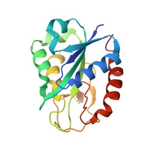
Flavodoxins, noncovalent complexes between apoflavodoxins and flavin mononucleotide (FMN), are useful models to investigate the mechanism of protein/flavin recognition. In this respect, the only available crystal structure of an apoflavodoxin (that from Anabaena) showed a closed isoalloxazine pocket and the presence of a bound phosphate ion, which posed many questions on the recognition mechanism and on the potential physiological role exerted by phosphate ions. To address these issues we report here the X-ray structure of the apoflavodoxin from the pathogen Helicobacter pylori. The protein naturally lacks one of the conserved aromatic residues that close the isoalloxazine pocket in Anabaena, and the structure has been determined in a medium lacking phosphate. In spite of these significant differences, the isoallozaxine pocket in H. pylori apoflavodoxin appears also closed and a chloride ion is bound at a native-like FMN phosphate site. It seems thus that it is a general characteristic of apoflavodoxins to display closed, non-native, isoalloxazine binding sites together with native-like, rather promiscuous, phosphate binding sites that can bear other available small anions present in solution. In this respect, both binding energy hot spots of the apoflavodoxin/FMN complex are initially unavailable to FMN binding and the specific spot for FMN recognition may depend on the dynamics of the two candidate regions. Molecular dynamics simulations show that the isoalloxazine binding loops are intrinsically flexible at physiological temperatures, thus facilitating the intercalation of the cofactor, and that their mobility is modulated by the anion bound at the phosphate site.
Organizational Affiliation:
Biocomputation and Complex Systems Physics Institute (BiFi), Universidad de Zaragoza, Unidad Asociada al IQFR-CSIC, Zaragoza, Spain.