2BMH

MODELING PROTEIN-SUBSTRATE INTERACTIONS IN THE HEME DOMAIN OF CYTOCHROME P450BM-3


Experimental Data Snapshot

  • Method: X-RAY DIFFRACTION
  • Resolution: 2.00 Å
  • R-Value Work: 0.184 
  • R-Value Observed: 0.184 

wwPDB Validation   3D Report Full Report


Ligand Structure Quality Assessment 


This is version 1.3 of the entry. See complete history


Literature

Modeling protein-substrate interactions in the heme domain of cytochrome P450(BM-3).

Li, H.Poulos, T.L.

(1995) Acta Crystallogr D Biol Crystallogr 51: 21-32

  • DOI: https://doi.org/10.1107/S0907444994009194
  • Primary Citation of Related Structures:  
    2BMH

  • PubMed Abstract: 

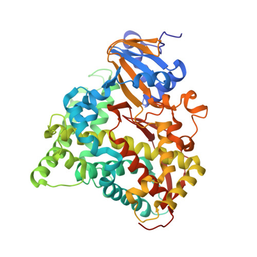
    The crystal structure of heme domain of the fatty acid monooxygenase, cytochrome P450(BM-3), consisting of residues 1-455 has been independently solved to R = 0.18 at 2.0 A. The crystal form used, space group P2(1) with two molecules per asymmetric unit, is isomorphous with that form with residues 1-471 first described by Boddupalli et al. [Boddupalli, Hasemann, Ravinchandran, Lu, Goldsmith, Deisenhofer & Peterson (1992). Proc. Natl Acad. Sci. USA, 89, 5567-5571] and used by Ravichandran, Boddupalli, Hasemann, Peterson & Deisenhofer [(1993). Science, 261, 731-736] to determine the crystal structure. The substrate-access channel consists of a large, hydrophobic cleft that appears to be the most likely route taken by fatty acid substrates. Attempts to soak crystals in mother liquor containing a variety of fatty acid substrates yielded featureless difference Fouriers even though fatty acid substrates are known to bind with dissociation constants in the micro M range. Modeling substrate-enzyme interactions reveals few contacts between the enzyme and substrate. More detailed modeling was carried out by subjecting both molecules in the asymmetric unit to extensive energy minimization. These studies reveal that the heme-domain active-site cleft can undergo a large conformational change that closes the access channel thereby providing enhanced protein-substrate interactions. These conformational changes are prevented from occurring by intermolecular contacts in the crystal lattice which lock the protein in the 'open' conformation.


  • Organizational Affiliation

    Department of Molecular Biology and Biochemistry, University of California at Irvine, 92717, USA.


Macromolecules
Find similar proteins by:  (by identity cutoff)  |  3D Structure
Entity ID: 1
MoleculeChains Sequence LengthOrganismDetailsImage
CYTOCHROME P450 BM-3
A, B
455Priestia megateriumMutation(s): 0 
EC: 1.14.14.1
UniProt
Find proteins for P14779 (Priestia megaterium (strain ATCC 14581 / DSM 32 / CCUG 1817 / JCM 2506 / NBRC 15308 / NCIMB 9376 / NCTC 10342 / NRRL B-14308 / VKM B-512 / Ford 19))
Explore P14779 
Go to UniProtKB:  P14779
Entity Groups  
Sequence Clusters30% Identity50% Identity70% Identity90% Identity95% Identity100% Identity
UniProt GroupP14779
Sequence Annotations
Expand
  • Reference Sequence
Small Molecules
Ligands 1 Unique
IDChains Name / Formula / InChI Key2D Diagram3D Interactions
HEM
Query on HEM

Download Ideal Coordinates CCD File 
C [auth A],
D [auth B]
PROTOPORPHYRIN IX CONTAINING FE
C34 H32 Fe N4 O4
KABFMIBPWCXCRK-RGGAHWMASA-L
Experimental Data & Validation

Experimental Data

  • Method: X-RAY DIFFRACTION
  • Resolution: 2.00 Å
  • R-Value Work: 0.184 
  • R-Value Observed: 0.184 
  • Space Group: P 1 21 1
Unit Cell:
Length ( Å )Angle ( ˚ )
a = 59.53α = 90
b = 154.03β = 94.97
c = 62.43γ = 90
Software Package:
Software NamePurpose
X-PLORmodel building
X-PLORrefinement
X-PLORphasing

Structure Validation

View Full Validation Report


Ligand Structure Quality Assessment 


Entry History 

Deposition Data

Revision History  (Full details and data files)

  • Version 1.0: 1994-07-31
    Type: Initial release
  • Version 1.1: 2008-03-24
    Changes: Version format compliance
  • Version 1.2: 2011-07-13
    Changes: Version format compliance
  • Version 1.3: 2024-02-14
    Changes: Data collection, Database references, Derived calculations, Other