2BBD

Crystal Structure of the STIV MCP


Experimental Data Snapshot

  • Method: X-RAY DIFFRACTION
  • Resolution: 2.04 Å
  • R-Value Free: 0.246 
  • R-Value Work: 0.207 
  • R-Value Observed: 0.207 

wwPDB Validation   3D Report Full Report


This is version 1.3 of the entry. See complete history


Literature

Structure of an archaeal virus capsid protein reveals a common ancestry to eukaryotic and bacterial viruses.

Khayat, R.Tang, L.Larson, E.T.Lawrence, C.M.Young, M.Johnson, J.E.

(2005) Proc Natl Acad Sci U S A 102: 18944-18949

  • DOI: https://doi.org/10.1073/pnas.0506383102
  • Primary Citation of Related Structures:  
    2BBD

  • PubMed Abstract: 

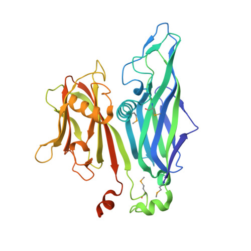
    Archaea and their viruses are poorly understood when compared with the Eukarya and Bacteria domains of life. We report here the crystal structure of the major capsid protein (MCP) of the Sulfolobus turreted icosahedral virus, an archaeal virus isolated from an acidic hot spring (pH 2-4, 72-92 degrees C) in Yellowstone National Park. The structure is nearly identical to the MCP structures of the eukaryotic Paramecium bursaria Chlorella virus, and the bacteriophage PRD1, and shows a common fold with the mammalian adenovirus. Structural analysis of the capsid architecture, determined by fitting the subunit into the electron cryomicroscopy reconstruction of the virus, identified a number of key interactions that are akin to those observed in adenovirus and PRD1. The similar capsid proteins and capsid architectures strongly suggest that these viral capsids originated and evolved from a common ancestor. Hence, this work provides a previously undescribed example of a viral relationship spanning the three domains of life (Eukarya, Bacteria, and Archaea). The MCP structure also provides insights into the stabilizing forces required for extracellular hyperthermophilic proteins to tolerate high-temperature hot springs.


  • Organizational Affiliation

    Department of Molecular Biology, The Scripps Research Institute, La Jolla, CA 92037, USA.


Macromolecules
Find similar proteins by:  (by identity cutoff)  |  3D Structure
Entity ID: 1
MoleculeChains Sequence LengthOrganismDetailsImage
coat protein
A, B, C, D
350Sulfolobus turreted icosahedral virus 1Mutation(s): 4 
UniProt
Find proteins for Q6Q0J0 (Sulfolobus turreted icosahedral virus 1)
Explore Q6Q0J0 
Go to UniProtKB:  Q6Q0J0
Entity Groups  
Sequence Clusters30% Identity50% Identity70% Identity90% Identity95% Identity100% Identity
UniProt GroupQ6Q0J0
Sequence Annotations
Expand
  • Reference Sequence
Small Molecules
Ligands 1 Unique
IDChains Name / Formula / InChI Key2D Diagram3D Interactions
MPD
Query on MPD

Download Ideal Coordinates CCD File 
E [auth A]
F [auth A]
G [auth A]
H [auth A]
I [auth A]
E [auth A],
F [auth A],
G [auth A],
H [auth A],
I [auth A],
J [auth A],
K [auth A],
L [auth A],
M [auth B],
N [auth C],
O [auth C],
P [auth C],
Q [auth C],
R [auth C],
S [auth D],
T [auth D],
U [auth D]
(4S)-2-METHYL-2,4-PENTANEDIOL
C6 H14 O2
SVTBMSDMJJWYQN-YFKPBYRVSA-N
Modified Residues  1 Unique
IDChains TypeFormula2D DiagramParent
MSE
Query on MSE
A, B, C, D
L-PEPTIDE LINKINGC5 H11 N O2 SeMET
Experimental Data & Validation

Experimental Data

  • Method: X-RAY DIFFRACTION
  • Resolution: 2.04 Å
  • R-Value Free: 0.246 
  • R-Value Work: 0.207 
  • R-Value Observed: 0.207 
  • Space Group: C 1 2 1
Unit Cell:
Length ( Å )Angle ( ˚ )
a = 241.6α = 90
b = 82.89β = 116
c = 114.8γ = 90
Software Package:
Software NamePurpose
CNSrefinement
DENZOdata reduction
SCALEPACKdata scaling
SnBphasing

Structure Validation

View Full Validation Report


Entry History 

Deposition Data

  • Released Date: 2005-12-06 
  • Deposition Author(s): Khayat, R.

Revision History  (Full details and data files)

  • Version 1.0: 2005-12-06
    Type: Initial release
  • Version 1.1: 2008-05-01
    Changes: Version format compliance
  • Version 1.2: 2011-07-13
    Changes: Derived calculations, Version format compliance
  • Version 1.3: 2017-10-18
    Changes: Refinement description