2B8T

Crystal structure of Thymidine Kinase from U.urealyticum in complex with thymidine


Experimental Data Snapshot

  • Method: X-RAY DIFFRACTION
  • Resolution: 2.00 Å
  • R-Value Free: 0.235 
  • R-Value Work: 0.195 
  • R-Value Observed: 0.197 

wwPDB Validation   3D Report Full Report


Ligand Structure Quality Assessment 


This is version 1.5 of the entry. See complete history


Literature

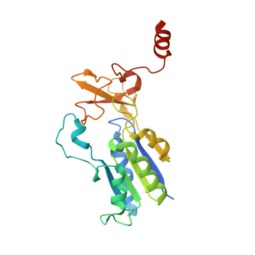
Structure of the substrate complex of thymidine kinase from Ureaplasma urealyticum and investigations of possible drug targets for the enzyme

Kosinska, U.Carnrot, C.Eriksson, S.Wang, L.Eklund, H.

(2005) FEBS Lett 272: 6365-6372

  • DOI: https://doi.org/10.1111/j.1742-4658.2005.05030.x
  • Primary Citation of Related Structures:  
    2B8T

  • PubMed Abstract: 

    Thymidine kinases have been found in most organisms, from viruses and bacteria to mammals. Ureaplasma urealyticum (parvum), which belongs to the class of cell-wall-lacking Mollicutes, has no de novo synthesis of DNA precursors and therefore has to rely on the salvage pathway. Thus, thymidine kinase (Uu-TK) is the key enzyme in dTTP synthesis. Recently the 3D structure of Uu-TK was determined in a feedback inhibitor complex, demonstrating that a lasso-like loop binds the thymidine moiety of the feedback inhibitor by hydrogen bonding to main-chain atoms. Here the structure with the substrate deoxythymidine is presented. The substrate binds similarly to the deoxythymidine part of the feedback inhibitor, and the lasso-like loop binds the base and deoxyribose moieties as in the complex determined previously. The catalytic base, Glu97, has a different position in the substrate complex from that in the complex with the feedback inhibitor, having moved in closer to the 5'-OH of the substrate to form a hydrogen bond. The phosphorylation of and inhibition by several nucleoside analogues were investigated and are discussed in the light of the substrate binding pocket, in comparison with human TK1. Kinetic differences between Uu-TK and human TK1 were observed that may be explained by structural differences. The tight interaction with the substrate allows minor substitutions at the 3 and 5 positions of the base, only fluorine substitutions at the 2'-Ara position, but larger substitutions at the 3' position of the deoxyribose.


  • Organizational Affiliation

    Department of Molecular Biology, Swedish University of Agricultural Sciences, Uppsala Biomedical Centre, Sweden.


Macromolecules
Find similar proteins by:  (by identity cutoff)  |  3D Structure
Entity ID: 1
MoleculeChains Sequence LengthOrganismDetailsImage
Thymidine kinase
A, B, C, D
223Ureaplasma parvumMutation(s): 1 
Gene Names: tdk
EC: 2.7.1.21
UniProt
Find proteins for Q9PPP5 (Ureaplasma parvum serovar 3 (strain ATCC 700970))
Explore Q9PPP5 
Go to UniProtKB:  Q9PPP5
Entity Groups  
Sequence Clusters30% Identity50% Identity70% Identity90% Identity95% Identity100% Identity
UniProt GroupQ9PPP5
Sequence Annotations
Expand
  • Reference Sequence
Small Molecules
Ligands 3 Unique
IDChains Name / Formula / InChI Key2D Diagram3D Interactions
THM
Query on THM

Download Ideal Coordinates CCD File 
F [auth A],
H [auth B],
J [auth C],
L [auth D]
THYMIDINE
C10 H14 N2 O5
IQFYYKKMVGJFEH-XLPZGREQSA-N
TRS
Query on TRS

Download Ideal Coordinates CCD File 
M [auth D]2-AMINO-2-HYDROXYMETHYL-PROPANE-1,3-DIOL
C4 H12 N O3
LENZDBCJOHFCAS-UHFFFAOYSA-O
ZN
Query on ZN

Download Ideal Coordinates CCD File 
E [auth A],
G [auth B],
I [auth C],
K [auth D]
ZINC ION
Zn
PTFCDOFLOPIGGS-UHFFFAOYSA-N
Experimental Data & Validation

Experimental Data

  • Method: X-RAY DIFFRACTION
  • Resolution: 2.00 Å
  • R-Value Free: 0.235 
  • R-Value Work: 0.195 
  • R-Value Observed: 0.197 
  • Space Group: P 1 21 1
Unit Cell:
Length ( Å )Angle ( ˚ )
a = 57.158α = 90
b = 115.649β = 101.02
c = 64.474γ = 90
Software Package:
Software NamePurpose
SCALAdata scaling
MOLREPphasing
REFMACrefinement
PDB_EXTRACTdata extraction
ProDCdata collection
CCP4data scaling

Structure Validation

View Full Validation Report


Ligand Structure Quality Assessment 


Entry History 

Deposition Data

Revision History  (Full details and data files)

  • Version 1.0: 2005-12-20
    Type: Initial release
  • Version 1.1: 2008-05-01
    Changes: Version format compliance
  • Version 1.2: 2011-07-13
    Changes: Version format compliance
  • Version 1.3: 2017-10-11
    Changes: Refinement description
  • Version 1.4: 2021-10-20
    Changes: Database references, Derived calculations
  • Version 1.5: 2023-08-23
    Changes: Data collection, Refinement description