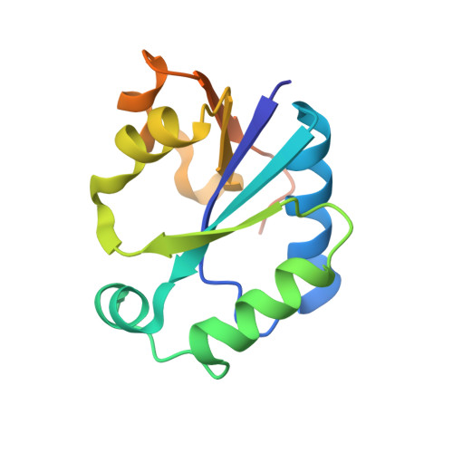
Crystal structure of CTP:glycerol-3-phosphate cytidylyltransferase from Staphylococcus aureus: examination of structural basis for kinetic mechanism.
Fong, D.H., Yim, V.C.-N., D'Elia, M.A., Brown, E.D., Berghuis, A.M.(2006) Biochim Biophys Acta 1764: 63-69
- PubMed: 16344011
- DOI: https://doi.org/10.1016/j.bbapap.2005.10.015
- Primary Citation of Related Structures:
2B7L - PubMed Abstract:
Integrity of the cell wall is essential for bacterial survival, and as a consequence components involved in its biosynthesis can potentially be exploited as targets for antibiotics. One such potential target is CTP:glycerol-3-phosphate cytidylyltransferase. This enzyme (TarD(Sa) in Staphylococcus aureus and TagD(Bs) in Bacillus subtilis) catalyzes the formation of CDP-glycerol, which is used for the assembly of linkages between peptidoglycan and teichoic acid polymer in Gram-positive bacteria. Intriguingly, despite the high sequence identity between TarD(Sa) and TagD(Bs) (69% identity), kinetic studies show that these two enzymes differ markedly in their kinetic mechanism and activity. To examine the basis for the disparate enzymological properties, we have determined the crystal structure of TarD(Sa) in the apo state to 3 A resolution, and performed equilibrium sedimentation analysis. Comparison of the structure with that of CTP- and CDP-glycerol-bound TagD(Bs) crystal structures reveals that the overall structure of TarD(Sa) is essentially the same as that of TagD(Bs), except in the C-terminus, where it forms a helix in TagD(Bs) but is disordered in the apo TarD(Sa) structure. In addition, TarD(Sa) can exist both as a tetramer and as a dimer, unlike TagD(Bs), which is a dimer. These observations shed light on the structural basis for the differing kinetic characteristics between TarD(Sa) and TagD(Bs).
Organizational Affiliation:
Department of Biochemistry, McGill University, 3655 Promenade Sir William Osler, Montreal, Quebec, Canada H3G 1Y6.