2B50

Human Nuclear Receptor-Ligand Complex 2


Experimental Data Snapshot

  • Method: X-RAY DIFFRACTION
  • Resolution: 2.00 Å
  • R-Value Free: 0.249 
  • R-Value Work: 0.197 
  • R-Value Observed: 0.199 

wwPDB Validation   3D Report Full Report


Ligand Structure Quality Assessment 


This is version 1.4 of the entry. See complete history


Literature

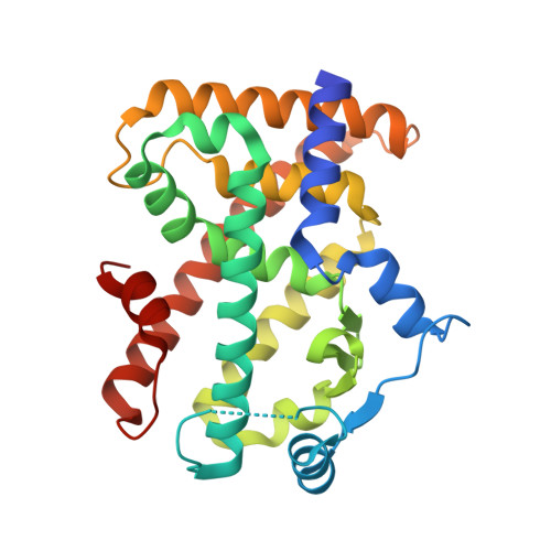
Recombinant Human PPAR-beta/delta Ligand-binding Domain is Locked in an Activated Conformation by Endogenous Fatty Acids

Fyffe, S.A.Alphey, M.S.Buetow, L.Smith, T.K.Ferguson, M.A.J.Sorensen, M.D.Bjorkling, F.Hunter, W.N.

(2006) J Mol Biol 356: 1005-1013

  • DOI: https://doi.org/10.1016/j.jmb.2005.12.047
  • Primary Citation of Related Structures:  
    2AWH, 2B50

  • PubMed Abstract: 

    High-resolution crystallographic structures of recombinant human peroxisome proliferator-activated receptor ligand-binding domain (isotype beta/delta) reveal a fatty acid in the binding site. Mass spectrometry confirmed the presence of C16:0, C16:1, C18:0 and C18:1 in a ratio of approximately 3:2:1:4 with 11, Z-octadecenoic acid (cis-vaccenic acid) identified as the predominant species. These are endogenous fatty acids acquired from the bacterial expression system, and serve to lock the ligand-binding domain into the activated conformation. A requirement for crystal growth, the additive n-heptyl-beta-d-glucopyranoside, binds near the activation function helix where recognition of co-activator proteins occurs. Our observations suggest potential physiological ligands for human PPAR-beta/delta and highlight that reported binding studies must be treated with caution unless endogenous fatty acids have been removed from the sample prior to analysis.


  • Organizational Affiliation

    Division of Biological Chemistry and Molecular Microbiology, School of Life Sciences, University of Dundee, Dundee DD1 5EH, UK.


Macromolecules
Find similar proteins by:  (by identity cutoff)  |  3D Structure
Entity ID: 1
MoleculeChains Sequence LengthOrganismDetailsImage
Peroxisome proliferator activated receptor delta
A, B
272Homo sapiensMutation(s): 0 
UniProt & NIH Common Fund Data Resources
Find proteins for Q03181 (Homo sapiens)
Explore Q03181 
Go to UniProtKB:  Q03181
PHAROS:  Q03181
GTEx:  ENSG00000112033 
Entity Groups  
Sequence Clusters30% Identity50% Identity70% Identity90% Identity95% Identity100% Identity
UniProt GroupQ03181
Sequence Annotations
Expand
  • Reference Sequence
Experimental Data & Validation

Experimental Data

  • Method: X-RAY DIFFRACTION
  • Resolution: 2.00 Å
  • R-Value Free: 0.249 
  • R-Value Work: 0.197 
  • R-Value Observed: 0.199 
  • Space Group: P 1 21 1
Unit Cell:
Length ( Å )Angle ( ˚ )
a = 39.776α = 90
b = 92.084β = 97
c = 95.017γ = 90
Software Package:
Software NamePurpose
REFMACrefinement
PDB_EXTRACTdata extraction
CCP4data scaling

Structure Validation

View Full Validation Report


Ligand Structure Quality Assessment 


Entry History 

Deposition Data

Revision History  (Full details and data files)

  • Version 1.0: 2006-02-14
    Type: Initial release
  • Version 1.1: 2008-05-01
    Changes: Version format compliance
  • Version 1.2: 2011-07-13
    Changes: Version format compliance
  • Version 1.3: 2020-07-29
    Type: Remediation
    Reason: Carbohydrate remediation
    Changes: Data collection, Database references, Derived calculations, Structure summary
  • Version 1.4: 2024-03-13
    Changes: Data collection, Database references, Structure summary