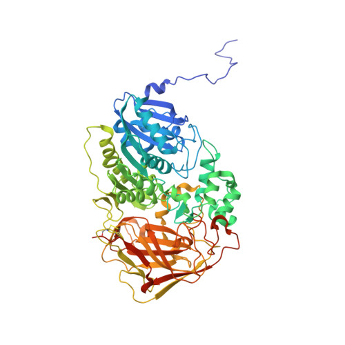
2B4K

Acetobacter turbidans alpha-amino acid ester hydrolase complexed with phenylglycine


Experimental Data Snapshot

  • Method: X-RAY DIFFRACTION
  • Resolution: 3.30 Å
  • R-Value Free: 0.289 
  • R-Value Work: 0.258 
  • R-Value Observed: 0.259 

wwPDB Validation   3D Report Full Report


Ligand Structure Quality Assessment 


This is version 1.4 of the entry. See complete history


Literature

Acetobacter turbidans alpha-amino acid ester hydrolase: how a single mutation improves an antibiotic-producing enzyme.

Barends, T.R.Polderman-Tijmes, J.J.Jekel, P.A.Williams, C.Wybenga, G.Janssen, D.B.Dijkstra, B.W.

(2006) J Biol Chem 281: 5804-5810

  • DOI: https://doi.org/10.1074/jbc.M511187200
  • Primary Citation of Related Structures:  
    1NX9, 1RYY, 2B4K, 2B9V

  • PubMed Abstract: 

    The alpha-amino acid ester hydrolase (AEH) from Acetobacter turbidans is a bacterial enzyme catalyzing the hydrolysis and synthesis of beta-lactam antibiotics. The crystal structures of the native enzyme, both unliganded and in complex with the hydrolysis product D-phenylglycine are reported, as well as the structures of an inactive mutant (S205A) complexed with the substrate ampicillin, and an active site mutant (Y206A) with an increased tendency to catalyze antibiotic production rather than hydrolysis. The structure of the native enzyme shows an acyl binding pocket, in which D-phenylglycine binds, and an additional space that is large enough to accommodate the beta-lactam moiety of an antibiotic. In the S205A mutant, ampicillin binds in this pocket in a non-productive manner, making extensive contacts with the side chain of Tyr(112), which also participates in oxyanion hole formation. In the Y206A mutant, the Tyr(112) side chain has moved with its hydroxyl group toward the catalytic serine. Because this changes the properties of the beta-lactam binding site, this could explain the increased beta-lactam transferase activity of this mutant.


  • Organizational Affiliation

    Laboratories of Biophysical Chemistry and Biochemistry, University of Groningen, Nijenborgh 4, 9747 AG Groningen, The Netherlands.


Macromolecules
Find similar proteins by:  (by identity cutoff)  |  3D Structure
Entity ID: 1
MoleculeChains Sequence LengthOrganismDetailsImage
Alpha-amino acid ester hydrolase
A, B, C, D
652Acetobacter pasteurianusMutation(s): 0 
Gene Names: aehA
UniProt
Find proteins for Q8VRK8 (Acetobacter pasteurianus)
Explore Q8VRK8 
Go to UniProtKB:  Q8VRK8
Entity Groups  
Sequence Clusters30% Identity50% Identity70% Identity90% Identity95% Identity100% Identity
UniProt GroupQ8VRK8
Sequence Annotations
Expand
  • Reference Sequence
Experimental Data & Validation

Experimental Data

  • Method: X-RAY DIFFRACTION
  • Resolution: 3.30 Å
  • R-Value Free: 0.289 
  • R-Value Work: 0.258 
  • R-Value Observed: 0.259 
  • Space Group: I 2 3
Unit Cell:
Length ( Å )Angle ( ˚ )
a = 340.909α = 90
b = 340.909β = 90
c = 340.909γ = 90
Software Package:
Software NamePurpose
REFMACrefinement
DENZOdata reduction
SCALEPACKdata scaling
MOLREPphasing

Structure Validation

View Full Validation Report


Ligand Structure Quality Assessment 


Entry History 

Deposition Data

Revision History  (Full details and data files)

  • Version 1.0: 2005-12-27
    Type: Initial release
  • Version 1.1: 2007-10-24
    Changes: Version format compliance
  • Version 1.2: 2011-07-13
    Changes: Derived calculations, Version format compliance
  • Version 1.3: 2022-12-21
    Changes: Database references, Derived calculations, Source and taxonomy, Structure summary
  • Version 1.4: 2023-09-20
    Changes: Data collection, Refinement description