2B3Y

Structure of a monoclinic crystal form of human cytosolic aconitase (IRP1)


Experimental Data Snapshot

  • Method: X-RAY DIFFRACTION
  • Resolution: 1.85 Å
  • R-Value Free: 0.261 
  • R-Value Work: 0.211 
  • R-Value Observed: 0.213 

wwPDB Validation   3D Report Full Report


This is version 1.3 of the entry. See complete history


Literature

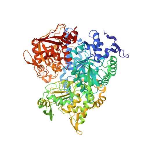
Crystal structure of human iron regulatory protein 1 as cytosolic aconitase

Dupuy, J.Volbeda, A.Carpentier, P.Darnault, C.Moulis, J.M.Fontecilla-Camps, J.C.

(2006) Structure 14: 129-139

  • DOI: https://doi.org/10.1016/j.str.2005.09.009
  • Primary Citation of Related Structures:  
    2B3X, 2B3Y

  • PubMed Abstract: 

    Iron regulatory proteins (IRPs) control the translation of proteins involved in iron uptake, storage and utilization by binding to specific noncoding sequences of the corresponding mRNAs known as iron-responsive elements (IREs). This strong interaction assures proper iron homeostasis in animal cells under iron shortage. Conversely, under iron-replete conditions, IRP1 binds a [4Fe-4S] cluster and functions as cytosolic aconitase. Regulation of the balance between the two IRP1 activities is complex, and it does not depend only on iron availability. Here, we report the crystal structure of human IRP1 in its aconitase form. Comparison with known structures of homologous enzymes reveals well-conserved folds and active site environments with significantly different surface shapes and charge distributions. The specific features of human IRP1 allow us to propose a tentative model of an IRP1-IRE complex that agrees with a range of previously obtained data.


  • Organizational Affiliation

    Laboratoire de Cristallographie et de Cristallogenèse des Protéines, Institut de Biologie Structurale JP Ebel, CEA/CNRS/Université Joseph Fourier, 38027 Grenoble Cedex 1, France.


Macromolecules
Find similar proteins by:  (by identity cutoff)  |  3D Structure
Entity ID: 1
MoleculeChains Sequence LengthOrganismDetailsImage
Iron-responsive element binding protein 1
A, B
888Homo sapiensMutation(s): 0 
EC: 4.2.1.3
UniProt & NIH Common Fund Data Resources
Find proteins for P21399 (Homo sapiens)
Explore P21399 
Go to UniProtKB:  P21399
PHAROS:  P21399
GTEx:  ENSG00000122729 
Entity Groups  
Sequence Clusters30% Identity50% Identity70% Identity90% Identity95% Identity100% Identity
UniProt GroupP21399
Sequence Annotations
Expand
  • Reference Sequence
Small Molecules
Ligands 4 Unique
IDChains Name / Formula / InChI Key2D Diagram3D Interactions
SF4
Query on SF4

Download Ideal Coordinates CCD File 
D [auth A],
I [auth B]
IRON/SULFUR CLUSTER
Fe4 S4
LJBDFODJNLIPKO-UHFFFAOYSA-N
GOL
Query on GOL

Download Ideal Coordinates CCD File 
E [auth A],
F [auth A]
GLYCEROL
C3 H8 O3
PEDCQBHIVMGVHV-UHFFFAOYSA-N
ACT
Query on ACT

Download Ideal Coordinates CCD File 
C [auth A],
G [auth B],
H [auth B]
ACETATE ION
C2 H3 O2
QTBSBXVTEAMEQO-UHFFFAOYSA-M
FMT
Query on FMT

Download Ideal Coordinates CCD File 
J [auth B]FORMIC ACID
C H2 O2
BDAGIHXWWSANSR-UHFFFAOYSA-N
Experimental Data & Validation

Experimental Data

  • Method: X-RAY DIFFRACTION
  • Resolution: 1.85 Å
  • R-Value Free: 0.261 
  • R-Value Work: 0.211 
  • R-Value Observed: 0.213 
  • Space Group: P 1 21 1
Unit Cell:
Length ( Å )Angle ( ˚ )
a = 63.91α = 90
b = 215.47β = 72
c = 64.01γ = 90
Software Package:
Software NamePurpose
REFMACrefinement
PDB_EXTRACTdata extraction
XDSdata reduction
XDSdata scaling
PHASERphasing

Structure Validation

View Full Validation Report


Entry History 

Deposition Data

Revision History  (Full details and data files)

  • Version 1.0: 2006-01-10
    Type: Initial release
  • Version 1.1: 2008-05-01
    Changes: Version format compliance
  • Version 1.2: 2011-07-13
    Changes: Advisory, Refinement description, Version format compliance
  • Version 1.3: 2023-08-23
    Changes: Data collection, Database references, Derived calculations, Refinement description