2B12

Crystal structure of the protein-protein complex between F82Y cytochrome c and cytochrome c peroxidase


Experimental Data Snapshot

  • Method: X-RAY DIFFRACTION
  • Resolution: 3.02 Å
  • R-Value Free: 0.268 
  • R-Value Work: 0.277 
  • R-Value Observed: 0.277 

wwPDB Validation   3D Report Full Report


Ligand Structure Quality Assessment 


This is version 2.1 of the entry. See complete history


Literature

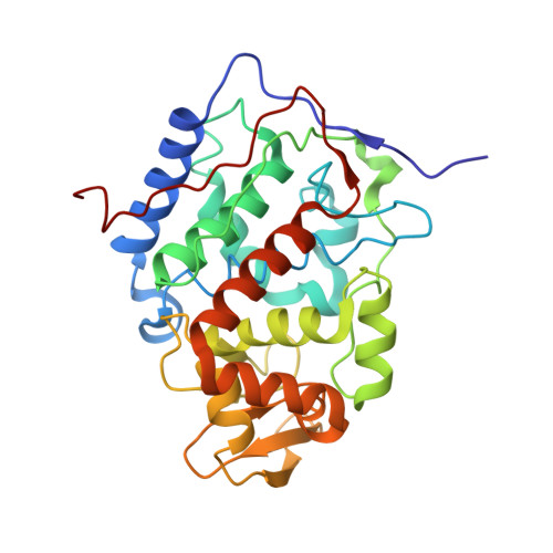
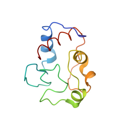
Effects of interface mutations on association modes and electron-transfer rates between proteins

Kang, S.A.Crane, B.R.

(2005) Proc Natl Acad Sci U S A 102: 15465-15470

  • DOI: https://doi.org/10.1073/pnas.0505176102
  • Primary Citation of Related Structures:  
    2B0Z, 2B10, 2B11, 2B12

  • PubMed Abstract: 

    Although bonding networks determine electron-transfer (ET) rates within proteins, the mechanism by which structure and dynamics influence ET across protein interfaces is not well understood. Measurements of photochemically induced ET and subsequent charge recombination between Zn-porphyrin-substituted cytochrome c peroxidase and cytochrome c in single crystals correlate reactivity with defined structures for different association modes of the redox partners. Structures and ET rates in crystals are consistent with tryptophan oxidation mediating charge recombination reactions. Conservative mutations at the interface can drastically affect how the proteins orient and dispose redox centers. Whereas some configurations are ET inactive, the wild-type complex exhibits the fastest recombination rate. Other association modes generate ET rates that do not correlate with predictions based on cofactor separations or simple bonding pathways. Inhibition of photoinduced ET at <273 K indicates gating by small-amplitude dynamics, even within the crystal. Thus, different associations achieve states of similar reactivity, and within those states conformational fluctuations enable interprotein ET.


  • Organizational Affiliation

    Department of Chemistry and Chemical Biology, Cornell University, Ithaca, NY 14853, USA.


Macromolecules
Find similar proteins by:  (by identity cutoff)  |  3D Structure
Entity ID: 1
MoleculeChains Sequence LengthOrganismDetailsImage
Cytochrome c peroxidase, mitochondrial294Saccharomyces cerevisiaeMutation(s): 0 
Gene Names: CCP1CCPCPO
EC: 1.11.1.5
UniProt
Find proteins for P00431 (Saccharomyces cerevisiae (strain ATCC 204508 / S288c))
Explore P00431 
Go to UniProtKB:  P00431
Entity Groups  
Sequence Clusters30% Identity50% Identity70% Identity90% Identity95% Identity100% Identity
UniProt GroupP00431
Sequence Annotations
Expand
  • Reference Sequence
Find similar proteins by:  (by identity cutoff)  |  3D Structure
Entity ID: 2
MoleculeChains Sequence LengthOrganismDetailsImage
Cytochrome c iso-1108Saccharomyces cerevisiaeMutation(s): 1 
Gene Names: CYC1
UniProt
Find proteins for P00044 (Saccharomyces cerevisiae (strain ATCC 204508 / S288c))
Explore P00044 
Go to UniProtKB:  P00044
Entity Groups  
Sequence Clusters30% Identity50% Identity70% Identity90% Identity95% Identity100% Identity
UniProt GroupP00044
Sequence Annotations
Expand
  • Reference Sequence
Small Molecules
Ligands 2 Unique
IDChains Name / Formula / InChI Key2D Diagram3D Interactions
ZNH
Query on ZNH

Download Ideal Coordinates CCD File 
C [auth A]PROTOPORPHYRIN IX CONTAINING ZN
C34 H32 N4 O4 Zn
FUTVBRXUIKZACV-RGGAHWMASA-L
HEC
Query on HEC

Download Ideal Coordinates CCD File 
D [auth B]HEME C
C34 H34 Fe N4 O4
HXQIYSLZKNYNMH-LJNAALQVSA-N
Experimental Data & Validation

Experimental Data

  • Method: X-RAY DIFFRACTION
  • Resolution: 3.02 Å
  • R-Value Free: 0.268 
  • R-Value Work: 0.277 
  • R-Value Observed: 0.277 
  • Space Group: P 21 21 21
Unit Cell:
Length ( Å )Angle ( ˚ )
a = 43.789α = 90
b = 51.832β = 90
c = 183.645γ = 90
Software Package:
Software NamePurpose
DENZOdata reduction
SCALEPACKdata scaling
REFMACrefinement
PDB_EXTRACTdata extraction
HKL-2000data reduction
AMoREphasing

Structure Validation

View Full Validation Report


Ligand Structure Quality Assessment 


Entry History 

Deposition Data

Revision History  (Full details and data files)

  • Version 1.0: 2005-10-25
    Type: Initial release
  • Version 1.1: 2008-05-01
    Changes: Version format compliance
  • Version 1.2: 2011-07-13
    Changes: Advisory, Refinement description, Version format compliance
  • Version 1.3: 2017-10-11
    Changes: Refinement description
  • Version 2.0: 2021-03-03
    Changes: Advisory, Atomic model, Data collection, Derived calculations, Non-polymer description, Structure summary
  • Version 2.1: 2021-10-20
    Changes: Database references