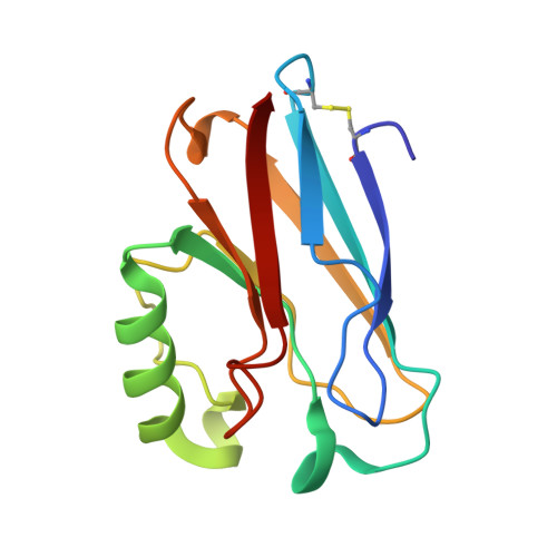
X-ray crystal structure of the two site-specific mutants His35Gln and His35Leu of azurin from Pseudomonas aeruginosa.
Nar, H., Messerschmidt, A., Huber, R., van de Kamp, M., Canters, G.W.(1991) J Mol Biol 218: 427-447
- PubMed: 1901363
- DOI: https://doi.org/10.1016/0022-2836(91)90723-j
- Primary Citation of Related Structures:
2AZU, 3AZU - PubMed Abstract:
The three-dimensional structures of two site-specific mutants of the blue copper protein azurin from Pseudomonas aeruginosa have been solved by a combination of isomorphous replacement and Patterson search techniques, and refined by energy-restrained least-squares methods. The mutations introduced by recombinant DNA techniques involve residue His35, which was exchanged for glutamine and leucine, to probe for its suggested role in electron transfer. The two mutants, His35Gln (H35Q) and His35Leu (H35L), crystallize non-isomorphously in the orthorhombic space group P2(1)2(1)2(1) with unit cell dimensions of a = 109.74 A, b = 99.15 A, c = 47.82 A for H35Q, a = 57.82 A, b = 81.06 A, c = 110.03 A for H35L. In each crystal form, there are four molecules in the asymmetric unit. They are arranged as a dimer of dimers in the H35Q case and are distorted from ideal C2 symmetry in H35L. The final crystallographic R-value is 16.3% for 20.747 reflections to a resolution of 2.1 A for H35Q and 17.0% for 32,548 reflections to 1.9 A for H35L. The crystal structures reported here represent the first crystallographically refined structures for azurin from P. aeruginosa. The structure is very similar to that of azurin from Alcaligenes denitrificans. The copper atom is located about 7 A below a hydrophobic surface region and is ligated by five donor groups in a distorted trigonal bipyramidal fashion. The implications for electron transfer properties of the protein are discussed in terms of the mutation site and the packing of the molecules within the tetramer.
Organizational Affiliation:
Max Planck Institut für Biochemie, Abteilung Strukturforschung Martinsried b. München, Germany.