2AZN

X-RAY Structure of 2,5-diamino-6-ribosylamino-4(3h)-pyrimidinone 5-phosphate reductase


Experimental Data Snapshot

  • Method: X-RAY DIFFRACTION
  • Resolution: 2.70 Å
  • R-Value Free: 0.253 
  • R-Value Work: 0.230 
  • R-Value Observed: 0.230 

wwPDB Validation   3D Report Full Report


Ligand Structure Quality Assessment 


This is version 1.2 of the entry. See complete history


Literature

Biosynthesis of riboflavin: structure and properties of 2,5-diamino-6-ribosylamino-4(3H)-pyrimidinone 5'-phosphate reductase of Methanocaldococcus jannaschii

Chatwell, L.Krojer, T.Fidler, A.Eisenreich, W.Bacher, A.Huber, R.Fischer, M.

(2006) J Mol Biol 359: 1334-1351

  • DOI: https://doi.org/10.1016/j.jmb.2006.04.045
  • Primary Citation of Related Structures:  
    2AZN

  • PubMed Abstract: 

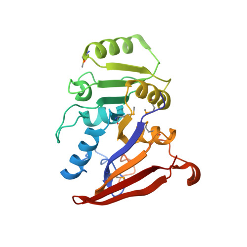
    The pyrimidine reductase of the riboflavin biosynthetic pathway (MjaRED) specified by the open reading frame MJ0671 of Methanocaldococcus jannaschii was expressed in Escherichia coli using a synthetic gene. The synthetic open reading frame that was optimized for expression in E. coli directed the synthesis of abundant amounts of the enzyme with an apparent subunit mass of 25 kDa. The enzyme was purified to apparent homogeneity and was shown to catalyze the conversion of 2,5-diamino-6-ribosylamino-4(3H)-pyrimidinone 5'-phosphate into 2,5-diamino-6-ribitylamino-4(3H)-pyrimidinone 5'-phosphate at a rate of 0.8 micromol min(-1) mg(-1) at pH 8.0 and at 30 degrees C. The protein is a homodimer as shown by sedimentation equilibrium analysis and sediments at an apparent velocity of 3.5 S. The structure of the enzyme in complex with the cofactor nicotinamide adenine dinucleotide phosphate was determined by X-ray crystallography at a resolution of 2.5 Angstroms. The folding pattern resembles that of dihydrofolate reductase with the Thermotoga maritima ortholog as the most similar structure. The substrate, 2,5-diamino-6-ribosylamino-4(3H)-pyrimidinone 5'-phosphate, was modeled into the putative active site. The model suggests the transfer of the pro-R hydrogen of C-4 of NADPH to C-1' of the substrate.


  • Organizational Affiliation

    Max-Planck Institut für Biochemie, Abteilung für Strukturforschung, Martinsried, Germany. chatwell@wzw.tum.de


Macromolecules
Find similar proteins by:  (by identity cutoff)  |  3D Structure
Entity ID: 1
MoleculeChains Sequence LengthOrganismDetailsImage
Putative 5-amino-6-(5-phosphoribosylamino)uracil reductase
A, B, C, D, E
A, B, C, D, E, F
219Methanocaldococcus jannaschiiMutation(s): 5 
EC: 1.1.1.193
UniProt
Find proteins for Q58085 (Methanocaldococcus jannaschii (strain ATCC 43067 / DSM 2661 / JAL-1 / JCM 10045 / NBRC 100440))
Explore Q58085 
Go to UniProtKB:  Q58085
Entity Groups  
Sequence Clusters30% Identity50% Identity70% Identity90% Identity95% Identity100% Identity
UniProt GroupQ58085
Sequence Annotations
Expand
  • Reference Sequence
Small Molecules
Ligands 3 Unique
IDChains Name / Formula / InChI Key2D Diagram3D Interactions
NAP
Query on NAP

Download Ideal Coordinates CCD File 
H [auth A]
K [auth B]
O [auth C]
Q [auth D]
R [auth E]
H [auth A],
K [auth B],
O [auth C],
Q [auth D],
R [auth E],
U [auth F]
NADP NICOTINAMIDE-ADENINE-DINUCLEOTIDE PHOSPHATE
C21 H28 N7 O17 P3
XJLXINKUBYWONI-NNYOXOHSSA-N
MA5
Query on MA5

Download Ideal Coordinates CCD File 
G [auth A]
I [auth B]
J [auth B]
M [auth C]
N [auth C]
G [auth A],
I [auth B],
J [auth B],
M [auth C],
N [auth C],
P [auth D],
S [auth F],
T [auth F]
2-(6-(2-CYCLOHEXYLETHOXY)-TETRAHYDRO-4,5-DIHYDROXY-2(HYDROXYMETHYL)-2H-PYRAN-3-YLOXY)-TETRAHYDRO-6(HYDROXYMETHYL)-2H-PY RAN-3,4,5-TRIOL
C20 H36 O11
RHXPDNGQJSXOMW-OIIXUNCGSA-N
EPE
Query on EPE

Download Ideal Coordinates CCD File 
L [auth B],
V [auth F]
4-(2-HYDROXYETHYL)-1-PIPERAZINE ETHANESULFONIC ACID
C8 H18 N2 O4 S
JKMHFZQWWAIEOD-UHFFFAOYSA-N
Modified Residues  1 Unique
IDChains TypeFormula2D DiagramParent
MSE
Query on MSE
A, B, C, D, E
A, B, C, D, E, F
L-PEPTIDE LINKINGC5 H11 N O2 SeMET
Experimental Data & Validation

Experimental Data

  • Method: X-RAY DIFFRACTION
  • Resolution: 2.70 Å
  • R-Value Free: 0.253 
  • R-Value Work: 0.230 
  • R-Value Observed: 0.230 
  • Space Group: P 43 21 2
Unit Cell:
Length ( Å )Angle ( ˚ )
a = 137.171α = 90
b = 137.171β = 90
c = 213.87γ = 90
Software Package:
Software NamePurpose
CNSrefinement

Structure Validation

View Full Validation Report


Ligand Structure Quality Assessment 


Entry History 

Deposition Data

Revision History  (Full details and data files)

  • Version 1.0: 2006-08-29
    Type: Initial release
  • Version 1.1: 2008-05-01
    Changes: Version format compliance
  • Version 1.2: 2011-07-13
    Changes: Derived calculations, Version format compliance