2AYL

2.0 Angstrom Crystal Structure of Manganese Protoporphyrin IX-reconstituted Ovine Prostaglandin H2 Synthase-1 Complexed With Flurbiprofen


Experimental Data Snapshot

  • Method: X-RAY DIFFRACTION
  • Resolution: 2.00 Å
  • R-Value Free: 0.237 
  • R-Value Work: 0.218 

wwPDB Validation   3D Report Full Report


Ligand Structure Quality Assessment 


This is version 2.1 of the entry. See complete history


Literature

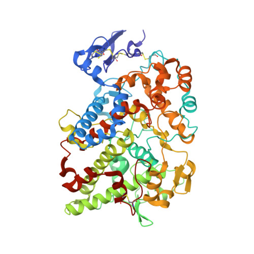
2.0 angstroms structure of prostaglandin H2 synthase-1 reconstituted with a manganese porphyrin cofactor.

Gupta, K.Selinsky, B.S.Loll, P.J.

(2006) Acta Crystallogr D Biol Crystallogr 62: 151-156

  • DOI: https://doi.org/10.1107/S0907444905036309
  • Primary Citation of Related Structures:  
    2AYL

  • PubMed Abstract: 

    Prostaglandin H2 synthase (EC 1.14.99.1) is a clinically important drug target that catalyzes two key steps in the biosynthesis of the eicosanoid hormones. The enzyme contains spatially distinct cyclooxygenase and peroxidase active sites, both of which require a heme cofactor. Substitution of ferric heme by Mn(III) protoporphyrin IX greatly diminishes the peroxidase activity, but has little effect on the cyclooxygenase activity. Here, the 2.0 angstroms resolution crystal structure of the Mn(III) form of ovine prostaglandin H2 synthase-1 is described (R = 21.8%, R(free) = 23.7%). Substitution of Mn(III) for Fe(III) causes no structural perturbations in the protein. However, the out-of-plane displacement of the manganese ion with respect to the porphyrin is greater than that of the iron by approximately 0.2 angstroms. This perturbation may help to explain the altered catalytic properties of the manganese enzyme.


  • Organizational Affiliation

    Department of Biochemistry and Molecular Biology, Drexel University College of Medicine, Philadelphia, PA 19102, USA.


Macromolecules
Find similar proteins by:  (by identity cutoff)  |  3D Structure
Entity ID: 1
MoleculeChains Sequence LengthOrganismDetailsImage
Prostaglandin G/H synthase 1
A, B
553Ovis ariesMutation(s): 0 
EC: 1.14.99.1
UniProt
Find proteins for P05979 (Ovis aries)
Explore P05979 
Go to UniProtKB:  P05979
Entity Groups  
Sequence Clusters30% Identity50% Identity70% Identity90% Identity95% Identity100% Identity
UniProt GroupP05979
Sequence Annotations
Expand
  • Reference Sequence
Oligosaccharides

Help

Entity ID: 2
MoleculeChains Length2D Diagram Glycosylation3D Interactions
2-acetamido-2-deoxy-beta-D-glucopyranose-(1-4)-2-acetamido-2-deoxy-beta-D-glucopyranose
C, F
2N-Glycosylation
Glycosylation Resources
GlyTouCan:  G42666HT
GlyCosmos:  G42666HT
GlyGen:  G42666HT
Entity ID: 3
MoleculeChains Length2D Diagram Glycosylation3D Interactions
alpha-D-mannopyranose-(1-6)-alpha-D-mannopyranose-(1-6)-alpha-D-mannopyranose-(1-4)-2-acetamido-2-deoxy-beta-D-glucopyranose-(1-4)-2-acetamido-2-deoxy-beta-D-glucopyranose
D, G
5N-Glycosylation
Glycosylation Resources
GlyTouCan:  G27747YT
GlyCosmos:  G27747YT
GlyGen:  G27747YT
Entity ID: 4
MoleculeChains Length2D Diagram Glycosylation3D Interactions
alpha-D-mannopyranose-(1-6)-alpha-D-mannopyranose-(1-4)-2-acetamido-2-deoxy-beta-D-glucopyranose-(1-4)-2-acetamido-2-deoxy-beta-D-glucopyranose
E, H
4N-Glycosylation
Glycosylation Resources
GlyTouCan:  G04854NQ
GlyCosmos:  G04854NQ
GlyGen:  G04854NQ
Small Molecules
Ligands 4 Unique
IDChains Name / Formula / InChI Key2D Diagram3D Interactions
MNH
Query on MNH

Download Ideal Coordinates CCD File 
N [auth A],
V [auth B]
MANGANESE PROTOPORPHYRIN IX
C34 H32 Mn N4 O4
UIBYYPDLEONCGQ-RGGAHWMASA-L
BOG
Query on BOG

Download Ideal Coordinates CCD File 
I [auth A]
J [auth A]
K [auth A]
L [auth A]
R [auth B]
I [auth A],
J [auth A],
K [auth A],
L [auth A],
R [auth B],
S [auth B],
T [auth B]
octyl beta-D-glucopyranoside
C14 H28 O6
HEGSGKPQLMEBJL-RKQHYHRCSA-N
FLP
Query on FLP

Download Ideal Coordinates CCD File 
M [auth A],
U [auth B]
FLURBIPROFEN
C15 H13 F O2
SYTBZMRGLBWNTM-JTQLQIEISA-N
GOL
Query on GOL

Download Ideal Coordinates CCD File 
O [auth A],
P [auth A],
Q [auth A],
W [auth B],
X [auth B]
GLYCEROL
C3 H8 O3
PEDCQBHIVMGVHV-UHFFFAOYSA-N
Binding Affinity Annotations 
IDSourceBinding Affinity
FLP BindingDB:  2AYL Ki: min: 75, max: 1000 (nM) from 3 assay(s)
IC50: min: 10, max: 2000 (nM) from 9 assay(s)
Experimental Data & Validation

Experimental Data

  • Method: X-RAY DIFFRACTION
  • Resolution: 2.00 Å
  • R-Value Free: 0.237 
  • R-Value Work: 0.218 
  • Space Group: I 2 2 2
Unit Cell:
Length ( Å )Angle ( ˚ )
a = 98.931α = 90
b = 206.55β = 90
c = 221.553γ = 90
Software Package:
Software NamePurpose
CNSrefinement
PDB_EXTRACTdata extraction
ADSCdata collection
d*TREKdata scaling
CNSphasing

Structure Validation

View Full Validation Report


Ligand Structure Quality Assessment 


Entry History 

Deposition Data

Revision History  (Full details and data files)

  • Version 1.0: 2006-01-24
    Type: Initial release
  • Version 1.1: 2008-05-01
    Changes: Version format compliance
  • Version 1.2: 2011-07-13
    Changes: Non-polymer description, Version format compliance
  • Version 2.0: 2020-07-29
    Type: Remediation
    Reason: Carbohydrate remediation
    Changes: Advisory, Atomic model, Data collection, Derived calculations, Structure summary
  • Version 2.1: 2023-08-23
    Changes: Data collection, Database references, Refinement description, Structure summary