2ATX

Crystal Structure of the TC10 GppNHp complex


Experimental Data Snapshot

  • Method: X-RAY DIFFRACTION
  • Resolution: 2.65 Å
  • R-Value Free: 0.260 
  • R-Value Work: 0.210 
  • R-Value Observed: 0.213 

wwPDB Validation   3D Report Full Report


Ligand Structure Quality Assessment 


This is version 1.5 of the entry. See complete history


Literature

An electrostatic steering mechanism of Cdc42 recognition by Wiskott-Aldrich syndrome proteins

Hemsath, L.Dvorsky, R.Fiegen, D.Carlier, M.F.Ahmadian, M.R.

(2005) Mol Cell 20: 313-324

  • DOI: https://doi.org/10.1016/j.molcel.2005.08.036
  • Primary Citation of Related Structures:  
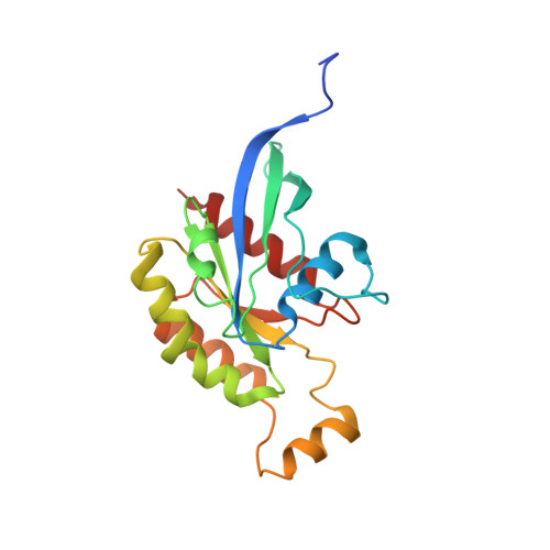
    2ATX

  • PubMed Abstract: 

    The specific and rapid formation of protein complexes is essential for diverse cellular processes such as remodeling of actin filaments in response to the interaction between Rho GTPases and the Wiskott-Aldrich syndrome proteins (WASp and N-WASp). Although Cdc42, TC10, and other members of the Rho family have been implicated in binding to and activating the WAS proteins, the exact nature of such a protein-protein recognition process has remained obscure. Here, we describe a mechanism that ensures rapid and selective long-range Cdc42-WASp recognition. The crystal structure of TC10, together with mutational and bioinformatic analyses, proved that the basic region of WASp and two unique glutamates in Cdc42 generate favorable electrostatic steering forces that control the accelerated WASp-Cdc42 association reaction. This process is a prerequisite for WASp activation and a critical step in temporal regulation and integration of WASp-mediated cellular responses.


  • Organizational Affiliation

    Abteilung Strukturelle Biologie, Max-Planck-Institut für Molekulare Physiologie, Otto-Hahn-Strasse 11, 44227 Dortmund, Germany.


Macromolecules
Find similar proteins by:  (by identity cutoff)  |  3D Structure
Entity ID: 1
MoleculeChains Sequence LengthOrganismDetailsImage
small GTP binding protein TC10
A, B
194Homo sapiensMutation(s): 0 
Gene Names: TC10
EC: 3.6.5.2
UniProt & NIH Common Fund Data Resources
Find proteins for P17081 (Homo sapiens)
Explore P17081 
Go to UniProtKB:  P17081
PHAROS:  P17081
GTEx:  ENSG00000119729 
Entity Groups  
Sequence Clusters30% Identity50% Identity70% Identity90% Identity95% Identity100% Identity
UniProt GroupP17081
Sequence Annotations
Expand
  • Reference Sequence
Experimental Data & Validation

Experimental Data

  • Method: X-RAY DIFFRACTION
  • Resolution: 2.65 Å
  • R-Value Free: 0.260 
  • R-Value Work: 0.210 
  • R-Value Observed: 0.213 
  • Space Group: P 21 21 21
Unit Cell:
Length ( Å )Angle ( ˚ )
a = 43.69α = 90
b = 82.2β = 90
c = 114.31γ = 90
Software Package:
Software NamePurpose
REFMACrefinement
ProDCdata collection
XDSdata scaling
MOLREPphasing

Structure Validation

View Full Validation Report


Ligand Structure Quality Assessment 


Entry History 

Deposition Data

Revision History  (Full details and data files)

  • Version 1.0: 2005-09-13
    Type: Initial release
  • Version 1.1: 2008-05-01
    Changes: Version format compliance
  • Version 1.2: 2011-07-13
    Changes: Advisory, Derived calculations, Version format compliance
  • Version 1.3: 2017-10-11
    Changes: Refinement description
  • Version 1.4: 2024-03-13
    Changes: Data collection, Database references, Derived calculations, Refinement description
  • Version 1.5: 2024-04-03
    Changes: Refinement description