2ATQ

RB69 single-stranded DNA binding protein-DNA polymerase fusion


Experimental Data Snapshot

  • Method: X-RAY DIFFRACTION
  • Resolution: 3.20 Å
  • R-Value Free: 0.345 
  • R-Value Work: 0.284 
  • R-Value Observed: 0.321 

wwPDB Validation   3D Report Full Report


Ligand Structure Quality Assessment 


This is version 1.5 of the entry. See complete history


Literature

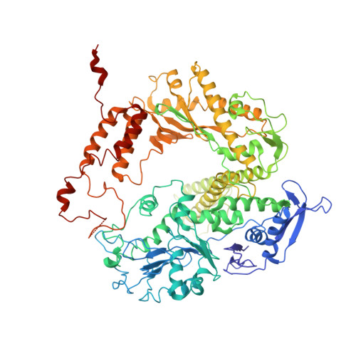
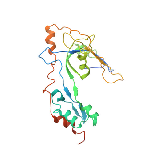
Structure and enzymatic properties of a chimeric bacteriophage RB69 DNA polymerase and single-stranded DNA binding protein with increased processivity.

Sun, S.Geng, L.Shamoo, Y.

(2006) Proteins 65: 231-238

  • DOI: https://doi.org/10.1002/prot.21088
  • Primary Citation of Related Structures:  
    2A1K, 2ATQ

  • PubMed Abstract: 

    In vivo, replicative DNA polymerases are made more processive by their interactions with accessory proteins at the replication fork. Single-stranded DNA binding protein (SSB) is an essential protein that binds tightly and cooperatively to single-stranded DNA during replication to remove adventitious secondary structures and protect the exposed DNA from endogenous nucleases. Using information from high resolution structures and biochemical data, we have engineered a functional chimeric enzyme of the bacteriophage RB69 DNA polymerase and SSB with substantially increased processivity. Fusion of RB69 DNA polymerase with its cognate SSB via a short six amino acid linker increases affinity for primer-template DNA by sixfold and subsequently increases processivity by sevenfold while maintaining fidelity. The crystal structure of this fusion protein was solved by a combination of multiwavelength anomalous diffraction and molecular replacement to 3.2 A resolution and shows that RB69 SSB is positioned proximal to the N-terminal domain of RB69 DNA polymerase near the template strand channel. The structural and biochemical data suggest that SSB interactions with DNA polymerase are transient and flexible, consistent with models of a dynamic replisome during elongation.


  • Organizational Affiliation

    Department of Biochemistry and Cell Biology, Rice University, Houston, Texas 77005, USA.


Macromolecules
Find similar proteins by:  (by identity cutoff)  |  3D Structure
Entity ID: 1
MoleculeChains Sequence LengthOrganismDetailsImage
DNA polymerase903Escherichia phage RB69Mutation(s): 2 
Gene Names: 43
EC: 2.7.7.7
UniProt
Find proteins for Q38087 (Escherichia phage RB69)
Explore Q38087 
Go to UniProtKB:  Q38087
Entity Groups  
Sequence Clusters30% Identity50% Identity70% Identity90% Identity95% Identity100% Identity
UniProt GroupQ38087
Sequence Annotations
Expand
  • Reference Sequence
Find similar proteins by:  (by identity cutoff)  |  3D Structure
Entity ID: 2
MoleculeChains Sequence LengthOrganismDetailsImage
gp32234Escherichia phage RB69Mutation(s): 0 
Gene Names: gp32
UniProt
Find proteins for Q7Y265 (Escherichia phage RB69)
Explore Q7Y265 
Go to UniProtKB:  Q7Y265
Entity Groups  
Sequence Clusters30% Identity50% Identity70% Identity90% Identity95% Identity100% Identity
UniProt GroupQ7Y265
Sequence Annotations
Expand
  • Reference Sequence
Small Molecules
Ligands 2 Unique
IDChains Name / Formula / InChI Key2D Diagram3D Interactions
GDP
Query on GDP

Download Ideal Coordinates CCD File 
C [auth A]GUANOSINE-5'-DIPHOSPHATE
C10 H15 N5 O11 P2
QGWNDRXFNXRZMB-UUOKFMHZSA-N
ZN
Query on ZN

Download Ideal Coordinates CCD File 
D [auth B]ZINC ION
Zn
PTFCDOFLOPIGGS-UHFFFAOYSA-N
Experimental Data & Validation

Experimental Data

  • Method: X-RAY DIFFRACTION
  • Resolution: 3.20 Å
  • R-Value Free: 0.345 
  • R-Value Work: 0.284 
  • R-Value Observed: 0.321 
  • Space Group: P 65
Unit Cell:
Length ( Å )Angle ( ˚ )
a = 196.029α = 90
b = 196.029β = 90
c = 85.17γ = 120
Software Package:
Software NamePurpose
DENZOdata reduction
SCALEPACKdata scaling
CNSrefinement
DMphasing
PDB_EXTRACTdata extraction
HKL-2000data reduction
CCP4data scaling
SHARPphasing

Structure Validation

View Full Validation Report


Ligand Structure Quality Assessment 


Entry History 

Deposition Data

Revision History  (Full details and data files)

  • Version 1.0: 2006-05-09
    Type: Initial release
  • Version 1.1: 2008-04-30
    Changes: Version format compliance
  • Version 1.2: 2011-07-13
    Changes: Version format compliance
  • Version 1.3: 2017-10-11
    Changes: Refinement description
  • Version 1.4: 2021-10-20
    Changes: Database references, Derived calculations
  • Version 1.5: 2023-08-23
    Changes: Data collection, Refinement description