Crystallographic and Pre-steady-state Kinetics Studies on Binding of NADH to Wild-type and Isoniazid-resistant Enoyl-ACP(CoA) Reductase Enzymes from Mycobacterium tuberculosis.
Oliveira, J.S., Pereira, J.H., Canduri, F., Rodrigues, N.C., de Souza, O.N., de Azevedo Jr., W.F., Basso, L.A., Santos, D.S.(2006) J Mol Biol 359: 646-666
- PubMed: 16647717
- DOI: https://doi.org/10.1016/j.jmb.2006.03.055
- Primary Citation of Related Structures:
2AQ8, 2AQH, 2AQI, 2AQK - PubMed Abstract:
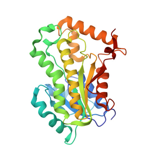
An understanding of isoniazid (INH) drug resistance mechanism in Mycobacterium tuberculosis should provide significant insight for the development of newer anti-tubercular agents able to control INH-resistant tuberculosis (TB). The inhA-encoded 2-trans enoyl-acyl carrier protein reductase enzyme (InhA) has been shown through biochemical and genetic studies to be the primary target for INH. In agreement with these results, mutations in the inhA structural gene have been found in INH-resistant clinical isolates of M.tuberculosis, the causative agent of TB. In addition, the InhA mutants were shown to have higher dissociation constant values for NADH and lower values for the apparent first-order rate constant for INH inactivation as compared to wild-type InhA. Here, in trying to identify structural changes between wild-type and INH-resistant InhA enzymes, we have solved the crystal structures of wild-type and of S94A, I47T and I21V InhA proteins in complex with NADH to resolutions of, respectively, 2.3A, 2.2A, 2.0 A, and 1.9A. The more prominent structural differences are located in, and appear to indirectly affect, the dinucleotide binding loop structure. Moreover, studies on pre-steady-state kinetics of NADH binding have been carried out. The results showed that the limiting rate constant values for NADH dissociation from the InhA-NADH binary complexes (k(off)) were eleven, five, and tenfold higher for, respectively, I21V, I47T, and S94A INH-resistant mutants of InhA as compared to INH-sensitive wild-type InhA. Accordingly, these results are proposed to be able to account for the reduction in affinity for NADH for the INH-resistant InhA enzymes.
Organizational Affiliation:
Centro de Pesquisas em Biologia Molecular e Funcional, Faculdade de Farmácia e Faculdade de Biociências, Instituto de Pesquisas Biomédicas, Pontifícia Universidade Católica do Rio Grande do Sul (PUCRS), Porto Alegre, RS 90619-900, Brasil.