2ANS

ADIPOCYTE LIPID BINDING PROTEIN COMPLEXED WITH 1-ANILINO-8-NAPHTHALENE SULFONATE


Experimental Data Snapshot

  • Method: X-RAY DIFFRACTION
  • Resolution: 2.50 Å
  • R-Value Free: 0.247 
  • R-Value Work: 0.189 
  • R-Value Observed: 0.189 

wwPDB Validation   3D Report Full Report


Ligand Structure Quality Assessment 


This is version 1.5 of the entry. See complete history


Literature

Studies of the ligand binding reaction of adipocyte lipid binding protein using the fluorescent probe 1, 8-anilinonaphthalene-8-sulfonate.

Ory, J.J.Banaszak, L.J.

(1999) Biophys J 77: 1107-1116

  • DOI: https://doi.org/10.1016/S0006-3495(99)76961-4
  • Primary Citation of Related Structures:  
    2ANS

  • PubMed Abstract: 

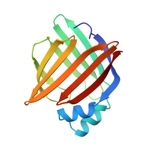
    The fluorescent probe anilinonaphthalene-8-sulfonate binds to adipocyte lipid binding protein at a site that competes with normal physiological ligands, such as fatty acids. Binding to the protein is accompanied by a relatively large increase in fluorescent intensity. To correlate the major change in optical properties and to determine the mechanism of competitive inhibition with fatty acids, the crystal structure of the protein with the bound fluorophore has been determined. In addition, the thermodynamic contributions to the binding reaction have been studied by titration calorimetry. Because the binding site is in a relatively internal position, kinetic studies have also been carried out to determine k(on). The results indicate that binding is not accompanied by any major conformational change. However, the negatively charged sulfonate moiety is not positioned the same as the carboxylate of fatty acid ligands as determined in previous studies. Nonetheless, the binding reaction is still driven by enthalpic effects. As judged by the crystallographic structure, a significant amount of the surface of the fluorophore is no longer exposed to water in the bound state.


  • Organizational Affiliation

    Department of Biochemistry, University of Minnesota, Minneapolis, Minnesota 55455, USA.


Macromolecules
Find similar proteins by:  (by identity cutoff)  |  3D Structure
Entity ID: 1
MoleculeChains Sequence LengthOrganismDetailsImage
ADIPOCYTE LIPID-BINDING PROTEIN
A, B
131Mus musculusMutation(s): 0 
UniProt
Find proteins for P04117 (Mus musculus)
Explore P04117 
Go to UniProtKB:  P04117
Entity Groups  
Sequence Clusters30% Identity50% Identity70% Identity90% Identity95% Identity100% Identity
UniProt GroupP04117
Sequence Annotations
Expand
  • Reference Sequence
Small Molecules
Binding Affinity Annotations 
IDSourceBinding Affinity
2AN BindingDB:  2ANS Ki: 1000 (nM) from 1 assay(s)
Kd: 150 (nM) from 1 assay(s)
Binding MOAD:  2ANS Kd: 1200 (nM) from 1 assay(s)
PDBBind:  2ANS Kd: 1200 (nM) from 1 assay(s)
Experimental Data & Validation

Experimental Data

  • Method: X-RAY DIFFRACTION
  • Resolution: 2.50 Å
  • R-Value Free: 0.247 
  • R-Value Work: 0.189 
  • R-Value Observed: 0.189 
  • Space Group: P 21 21 21
Unit Cell:
Length ( Å )Angle ( ˚ )
a = 80.8α = 90
b = 96.1β = 90
c = 49.9γ = 90
Software Package:
Software NamePurpose
X-PLORrefinement
X-PLORmodel building
CNSrefinement
XENGENdata reduction
XENGENdata scaling
CNSphasing
X-PLORphasing

Structure Validation

View Full Validation Report


Ligand Structure Quality Assessment 


Entry History 

Deposition Data

Revision History  (Full details and data files)

  • Version 1.0: 1999-07-22
    Type: Initial release
  • Version 1.1: 2008-03-24
    Changes: Version format compliance
  • Version 1.2: 2011-07-13
    Changes: Non-polymer description, Version format compliance
  • Version 1.3: 2019-07-17
    Changes: Data collection, Refinement description, Structure summary
  • Version 1.4: 2019-08-14
    Changes: Data collection
  • Version 1.5: 2023-08-09
    Changes: Database references, Derived calculations, Refinement description