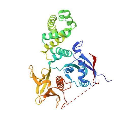
Crystal Structure of the FERM Domain of Focal Adhesion Kinase
Ceccarelli, D.F., Song, H.K., Poy, F., Schaller, M.D., Eck, M.J.(2006) J Biol Chem 281: 252-259
- PubMed: 16221668
- DOI: https://doi.org/10.1074/jbc.M509188200
- Primary Citation of Related Structures:
2AEH, 2AL6 - PubMed Abstract:
Focal adhesion kinase (FAK) is a non-receptor tyrosine kinase that localizes to focal adhesions in adherent cells. Through phosphorylation of proteins assembled at the cytoplasmic tails of integrins, FAK promotes signaling events that modulate cellular growth, survival, and migration. The amino-terminal region of FAK contains a region of sequence homology with band 4.1 and ezrin/radixin/moesin (ERM) proteins termed a FERM domain. FERM domains are found in a variety of signaling and cytoskeletal proteins and are thought to mediate intermolecular interactions with partner proteins and phospholipids at the plasma membrane and intramolecular regulatory interactions. Here we report two crystal structures of an NH2-terminal fragment of avian FAK containing the FERM domain and a portion of the regulatory linker that connects the FERM and kinase domains. The tertiary folds of the three subdomains (F1, F2, and F3) are similar to those of known FERM structures despite low sequence conservation. Differences in the sequence and relative orientation of the F3 subdomain alters the nature of the interdomain interface, and the phosphoinositide binding site found in ERM family FERM domains is not present in FAK. A putative protein interaction site on the F3 lobe is masked by the proximal region of the linker. Additionally, in one structure the adjacent Src SH3 and SH2 binding sites in the linker associate with the surfaces of the F3 and F1 lobes, respectively. These structural features suggest the possibility that protein interactions of the FAK FERM domain can be regulated by binding of Src kinases to the linker segment.
Organizational Affiliation:
Department of Cancer Biology, Dana-Farber Cancer Institute, Boston, Massachusetts 02115, USA.