2AL1

Crystal Structure Analysis of Enolase Mg Subunit Complex at pH 8.0


Experimental Data Snapshot

  • Method: X-RAY DIFFRACTION
  • Resolution: 1.50 Å
  • R-Value Free: 0.217 
  • R-Value Work: 0.153 
  • R-Value Observed: 0.153 

wwPDB Validation   3D Report Full Report


Ligand Structure Quality Assessment 


This is version 1.3 of the entry. See complete history


Literature

Structure and catalytic properties of an engineered heterodimer of enolase composed of one active and one inactive subunit

Sims, P.A.Menefee, A.L.Larsen, T.M.Mansoorabadi, S.O.Reed, G.H.

(2006) J Mol Biol 355: 422-431

  • DOI: https://doi.org/10.1016/j.jmb.2005.10.050
  • Primary Citation of Related Structures:  
    2AL1, 2AL2

  • PubMed Abstract: 

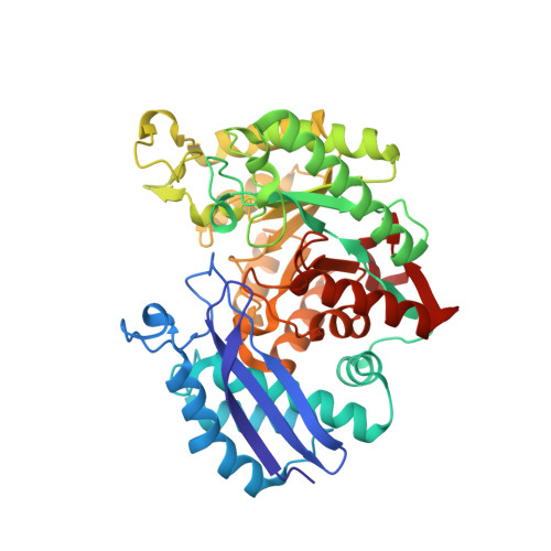
    Enolase is a dimeric enzyme that catalyzes the interconversion of 2-phospho-D-glycerate and phosphoenolpyruvate. This reversible dehydration is effected by general acid-base catalysis that involves, principally, Lys345 and Glu211 (numbering system of enolase 1 from yeast). The crystal structure of the inactive E211Q enolase shows that the protein is properly folded. However, K345 variants have, thus far, failed to crystallize. This problem was solved by crystallization of an engineered heterodimer of enolase. The heterodimer was composed of an inactive subunit that has a K345A mutation and an active subunit that has N80D and N126D surface mutations to facilitate ion-exchange chromatographic separation of the three dimeric species. The structure of this heterodimeric variant, in complex with substrate/product, was obtained at 1.85 A resolution. The structure was compared to a new structure of wild-type enolase obtained from crystals belonging to the same space group. Asymmetric dimers having one subunit exhibiting two of the three active site loops in an open conformation and the other in a conformation having all three loops closed appear in both structures. The K345A subunit of the heterodimer is in the loop-closed conformation; its Calpha carbon atoms closely match those of the corresponding subunit of wild-type enolase (root-mean-squared deviation of 0.23 A). The kcat and kcat/Km values of the heterodimer are approximately half those of the N80D/N126D homodimer, which suggests that the subunits in solution are kinetically independent. A comparison of enolase structures obtained from crystals belonging to different space groups suggests that asymmetric dimers can be a consequence of the asymmetric positioning of the subunits within the crystal lattice.


  • Organizational Affiliation

    Department of Biochemistry, University of Wisconsin-Madison, Madison, WI, 53726, USA.


Macromolecules
Find similar proteins by:  (by identity cutoff)  |  3D Structure
Entity ID: 1
MoleculeChains Sequence LengthOrganismDetailsImage
enolase 1
A, B
436Saccharomyces cerevisiaeMutation(s): 0 
EC: 4.2.1.11
UniProt
Find proteins for P00924 (Saccharomyces cerevisiae (strain ATCC 204508 / S288c))
Explore P00924 
Go to UniProtKB:  P00924
Entity Groups  
Sequence Clusters30% Identity50% Identity70% Identity90% Identity95% Identity100% Identity
UniProt GroupP00924
Sequence Annotations
Expand
  • Reference Sequence
Small Molecules
Ligands 5 Unique
IDChains Name / Formula / InChI Key2D Diagram3D Interactions
2PG
Query on 2PG

Download Ideal Coordinates CCD File 
F [auth A]2-PHOSPHOGLYCERIC ACID
C3 H7 O7 P
GXIURPTVHJPJLF-UWTATZPHSA-N
PEP
Query on PEP

Download Ideal Coordinates CCD File 
E [auth A],
L [auth B]
PHOSPHOENOLPYRUVATE
C3 H5 O6 P
DTBNBXWJWCWCIK-UHFFFAOYSA-N
K
Query on K

Download Ideal Coordinates CCD File 
J [auth B],
K [auth B]
POTASSIUM ION
K
NPYPAHLBTDXSSS-UHFFFAOYSA-N
CL
Query on CL

Download Ideal Coordinates CCD File 
I [auth B]CHLORIDE ION
Cl
VEXZGXHMUGYJMC-UHFFFAOYSA-M
MG
Query on MG

Download Ideal Coordinates CCD File 
C [auth A],
D [auth A],
G [auth B],
H [auth B]
MAGNESIUM ION
Mg
JLVVSXFLKOJNIY-UHFFFAOYSA-N
Experimental Data & Validation

Experimental Data

  • Method: X-RAY DIFFRACTION
  • Resolution: 1.50 Å
  • R-Value Free: 0.217 
  • R-Value Work: 0.153 
  • R-Value Observed: 0.153 
  • Space Group: P 1 21 1
Unit Cell:
Length ( Å )Angle ( ˚ )
a = 72α = 90
b = 65β = 99.5
c = 85.9γ = 90
Software Package:
Software NamePurpose
HKL-2000data collection
SCALEPACKdata scaling
RASMOLmodel building
SHELXL-97refinement
HKL-2000data reduction

Structure Validation

View Full Validation Report


Ligand Structure Quality Assessment 


Entry History 

Deposition Data

Revision History  (Full details and data files)

  • Version 1.0: 2006-01-24
    Type: Initial release
  • Version 1.1: 2008-04-30
    Changes: Version format compliance
  • Version 1.2: 2011-07-13
    Changes: Version format compliance
  • Version 1.3: 2024-02-14
    Changes: Data collection, Database references, Derived calculations