2AJC

Porcine dipeptidyl peptidase IV (CD26) in complex with 4-(2-Aminoethyl)-benzene sulphonyl fluoride (AEBSF)


Experimental Data Snapshot

  • Method: X-RAY DIFFRACTION
  • Resolution: 1.95 Å
  • R-Value Free: 0.234 
  • R-Value Work: 0.202 

wwPDB Validation   3D Report Full Report


Ligand Structure Quality Assessment 


This is version 2.1 of the entry. See complete history


Literature

Rigidity and flexibility of dipeptidyl peptidase IV: crystal structures of and docking experiments with DPIV.

Engel, M.Hoffmann, T.Manhart, S.Heiser, U.Chambre, S.Huber, R.Demuth, H.U.Bode, W.

(2006) J Mol Biol 355: 768-783

  • DOI: https://doi.org/10.1016/j.jmb.2005.11.014
  • Primary Citation of Related Structures:  
    2AJ8, 2AJB, 2AJC, 2AJD

  • PubMed Abstract: 

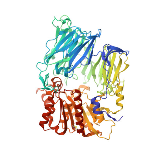
    Dipeptidyl peptidase IV (DPIV) is an alpha,beta-hydrolase-like serine exopeptidase, which removes dipeptides, preferentially with a C-terminal l-Pro residue, from the N terminus of longer peptide substrates. Previously, we determined the tetrameric 1.8A crystal structure of native porcine DPIV. Each monomer is composed of a beta-propeller and a catalytic domain, which together embrace an internal cavity housing the active centre. This cavity is connected to the bulk solvent by a "propeller opening" and a "side opening". Here, we analyse DPIV complexes with a t-butyl-Gly-Pro-Ile tripeptide, Pro-boroPro, a piperazine purine compound, and aminoethyl phenyl sulfonylfluoride. The latter two compounds bind to the active-site groove in a compact and a quite bulky manner, respectively, causing considerable shifts of the catalytic Ser630 side-chain and of the Tyr547 phenolic group, which forms the oxyanion hole. The tripeptide, mimicking a peptide substrate, is clamped to the active site through tight interactions via its N-terminal alpha-ammonium group, the P2 carbonyl group, the P1-l-Pro side-chain, the C-terminal carboxylate group, and the stable orthoacid ester amide formed between the scissile peptide carbonyl group and Ser630 O(gamma). This stable trapping of the tripeptide could be due to stabilization of the protonated His740 imidazolium cation by the adjacent negatively charged C-terminal carboxylate group, preventing proton transfer to the leaving group nitrogen atom. Docking experiments with the compact rigid 58 residue protein aprotinin, which had been shown to be processed by DPIV, indicate that the Arg1-Pro2 N terminus can access the DPIV active site only upon widening of its side openings, probably by separation of the first and the last propeller blades, and/or of the catalytic and the propeller domain.


  • Organizational Affiliation

    Max-Planck-Institut für Biochemie, AG Proteinaseforschung, Am Klopferspitz 18, D-82152 Martinsried, Germany.


Macromolecules
Find similar proteins by:  (by identity cutoff)  |  3D Structure
Entity ID: 1
MoleculeChains Sequence LengthOrganismDetailsImage
Dipeptidyl peptidase 4
A, B, C, D
728Sus scrofaMutation(s): 0 
EC: 3.4.14.5
UniProt
Find proteins for P22411 (Sus scrofa)
Explore P22411 
Go to UniProtKB:  P22411
Entity Groups  
Sequence Clusters30% Identity50% Identity70% Identity90% Identity95% Identity100% Identity
UniProt GroupP22411
Sequence Annotations
Expand
  • Reference Sequence
Oligosaccharides

Help

Entity ID: 2
MoleculeChains Length2D Diagram Glycosylation3D Interactions
2-acetamido-2-deoxy-beta-D-glucopyranose-(1-4)-2-acetamido-2-deoxy-beta-D-glucopyranose
E, F, H, I, K
E, F, H, I, K, L, M
2N-Glycosylation
Glycosylation Resources
GlyTouCan:  G42666HT
GlyCosmos:  G42666HT
GlyGen:  G42666HT
Entity ID: 3
MoleculeChains Length2D Diagram Glycosylation3D Interactions
beta-D-mannopyranose-(1-4)-2-acetamido-2-deoxy-beta-D-glucopyranose-(1-4)-2-acetamido-2-deoxy-beta-D-glucopyranose
G, J
3N-Glycosylation
Glycosylation Resources
GlyTouCan:  G15407YE
GlyCosmos:  G15407YE
GlyGen:  G15407YE
Small Molecules
Ligands 3 Unique
IDChains Name / Formula / InChI Key2D Diagram3D Interactions
NAG
Query on NAG

Download Ideal Coordinates CCD File 
AA [auth C]
BA [auth C]
CA [auth C]
DA [auth C]
GA [auth D]
AA [auth C],
BA [auth C],
CA [auth C],
DA [auth C],
GA [auth D],
HA [auth D],
N [auth A],
O [auth A],
P [auth A],
Q [auth A],
T [auth B],
U [auth B],
V [auth B],
W [auth B],
Z [auth C]
2-acetamido-2-deoxy-beta-D-glucopyranose
C8 H15 N O6
OVRNDRQMDRJTHS-FMDGEEDCSA-N
AES
Query on AES

Download Ideal Coordinates CCD File 
FA [auth C],
JA [auth D],
S [auth A],
Y [auth B]
4-(2-AMINOETHYL)BENZENESULFONYL FLUORIDE
C8 H10 F N O2 S
MGSKVZWGBWPBTF-UHFFFAOYSA-N
SO4
Query on SO4

Download Ideal Coordinates CCD File 
EA [auth C],
IA [auth D],
R [auth A],
X [auth B]
SULFATE ION
O4 S
QAOWNCQODCNURD-UHFFFAOYSA-L
Experimental Data & Validation

Experimental Data

  • Method: X-RAY DIFFRACTION
  • Resolution: 1.95 Å
  • R-Value Free: 0.234 
  • R-Value Work: 0.202 
  • Space Group: P 1
Unit Cell:
Length ( Å )Angle ( ˚ )
a = 62.35α = 112.76
b = 118.68β = 95.16
c = 133.68γ = 90.95
Software Package:
Software NamePurpose
DENZOdata reduction
SCALEPACKdata scaling
CNSrefinement
PDB_EXTRACTdata extraction
CNSphasing

Structure Validation

View Full Validation Report


Ligand Structure Quality Assessment 


Entry History 

Deposition Data

Revision History  (Full details and data files)

  • Version 1.0: 2006-02-28
    Type: Initial release
  • Version 1.1: 2008-04-30
    Changes: Version format compliance
  • Version 1.2: 2011-07-13
    Changes: Non-polymer description, Version format compliance
  • Version 1.3: 2012-06-06
    Changes: Non-polymer description
  • Version 1.4: 2017-10-11
    Changes: Advisory, Refinement description
  • Version 2.0: 2020-07-29
    Type: Remediation
    Reason: Carbohydrate remediation
    Changes: Atomic model, Data collection, Derived calculations, Structure summary
  • Version 2.1: 2023-08-23
    Changes: Advisory, Data collection, Database references, Refinement description, Structure summary