2AHX

Crystal structure of ErbB4/HER4 extracellular domain


Experimental Data Snapshot

  • Method: X-RAY DIFFRACTION
  • Resolution: 2.40 Å
  • R-Value Free: 0.265 
  • R-Value Work: 0.235 
  • R-Value Observed: 0.237 

wwPDB Validation   3D Report Full Report


Ligand Structure Quality Assessment 


This is version 2.0 of the entry. See complete history


Literature

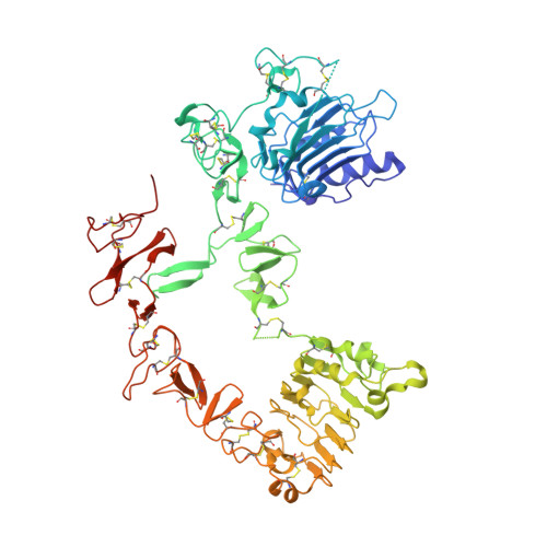
The extracellular region of ErbB4 adopts a tethered conformation in the absence of ligand

Bouyain, S.Longo, P.A.Li, S.Ferguson, K.M.Leahy, D.J.

(2005) Proc Natl Acad Sci U S A 102: 15024-15029

  • DOI: https://doi.org/10.1073/pnas.0507591102
  • Primary Citation of Related Structures:  
    2AHX

  • PubMed Abstract: 

    The human ErbB family of receptor tyrosine kinases comprises the epidermal growth factor receptor (EGFR/ErbB1/HER1), ErbB2 (HER2/Neu), ErbB3 (HER3), and ErbB4 (HER4). ErbBs play fundamental roles in cell growth and differentiation events in embryonic and adult tissues, and inappropriate ErbB activity has been implicated in several human cancers. We report here the 2.4 A crystal structure of the extracellular region of human ErbB4 in the absence of ligand and show that it adopts a tethered conformation similar to inactive forms of ErbB1 and ErbB3. This structure completes the gallery of unliganded ErbB receptors and demonstrates that all human ligand-binding ErbBs adopt the autoinhibited conformation. We also show that the binding of neuregulin-1beta to ErbB4 and ErbB3 and the binding of betacellulin to both ErbB4 and ErbB1 does not decrease at low pH, unlike the binding of epidermal growth factor and transforming growth factor-alpha to ErbB1. These results indicate an important role for ligand in determining pH-dependent binding and may explain different responses observed when the same ErbB receptor is stimulated by different ligands.


  • Organizational Affiliation

    Department of Biophysics and Biophysical Chemistry, Johns Hopkins University School of Medicine, Baltimore, MD 21205, USA.


Macromolecules
Find similar proteins by:  (by identity cutoff)  |  3D Structure
Entity ID: 1
MoleculeChains Sequence LengthOrganismDetailsImage
Receptor tyrosine-protein kinase erbB-4
A, B
617Homo sapiensMutation(s): 1 
Gene Names: ERBB4HER4
EC: 2.7.1.112
UniProt & NIH Common Fund Data Resources
Find proteins for Q15303 (Homo sapiens)
Explore Q15303 
Go to UniProtKB:  Q15303
PHAROS:  Q15303
GTEx:  ENSG00000178568 
Entity Groups  
Sequence Clusters30% Identity50% Identity70% Identity90% Identity95% Identity100% Identity
UniProt GroupQ15303
Sequence Annotations
Expand
  • Reference Sequence
Small Molecules
Ligands 3 Unique
IDChains Name / Formula / InChI Key2D Diagram3D Interactions
NAG
Query on NAG

Download Ideal Coordinates CCD File 
C [auth A]
D [auth A]
E [auth A]
F [auth A]
G [auth A]
C [auth A],
D [auth A],
E [auth A],
F [auth A],
G [auth A],
H [auth A],
I [auth A],
J [auth A],
L [auth B],
M [auth B],
N [auth B],
O [auth B],
P [auth B],
Q [auth B],
R [auth B],
S [auth B]
2-acetamido-2-deoxy-beta-D-glucopyranose
C8 H15 N O6
OVRNDRQMDRJTHS-FMDGEEDCSA-N
SO4
Query on SO4

Download Ideal Coordinates CCD File 
K [auth A],
T [auth B],
U [auth B]
SULFATE ION
O4 S
QAOWNCQODCNURD-UHFFFAOYSA-L
YT3
Query on YT3

Download Ideal Coordinates CCD File 
V [auth B]YTTRIUM (III) ION
Y
GRTBAGCGDOYUBE-UHFFFAOYSA-N
Experimental Data & Validation

Experimental Data

  • Method: X-RAY DIFFRACTION
  • Resolution: 2.40 Å
  • R-Value Free: 0.265 
  • R-Value Work: 0.235 
  • R-Value Observed: 0.237 
  • Space Group: C 2 2 21
Unit Cell:
Length ( Å )Angle ( ˚ )
a = 76.96α = 90
b = 203.09β = 90
c = 261.32γ = 90
Software Package:
Software NamePurpose
DENZOdata reduction
SCALEPACKdata scaling
AMoREphasing
REFMACrefinement

Structure Validation

View Full Validation Report


Ligand Structure Quality Assessment 


Entry History 

Deposition Data

Revision History  (Full details and data files)

  • Version 1.0: 2005-09-27
    Type: Initial release
  • Version 1.1: 2008-04-30
    Changes: Version format compliance
  • Version 1.2: 2011-07-13
    Changes: Advisory, Version format compliance
  • Version 1.3: 2017-10-11
    Changes: Refinement description
  • Version 1.4: 2018-03-14
    Changes: Database references
  • Version 2.0: 2020-07-29
    Type: Remediation
    Reason: Carbohydrate remediation
    Changes: Advisory, Atomic model, Data collection, Database references, Derived calculations, Non-polymer description, Structure summary