2AHE

Crystal structure of a soluble form of CLIC4. intercellular chloride ion channel


Experimental Data Snapshot

  • Method: X-RAY DIFFRACTION
  • Resolution: 1.80 Å
  • R-Value Free: 0.229 
  • R-Value Work: 0.194 
  • R-Value Observed: 0.196 

wwPDB Validation   3D Report Full Report


This is version 1.3 of the entry. See complete history


Literature

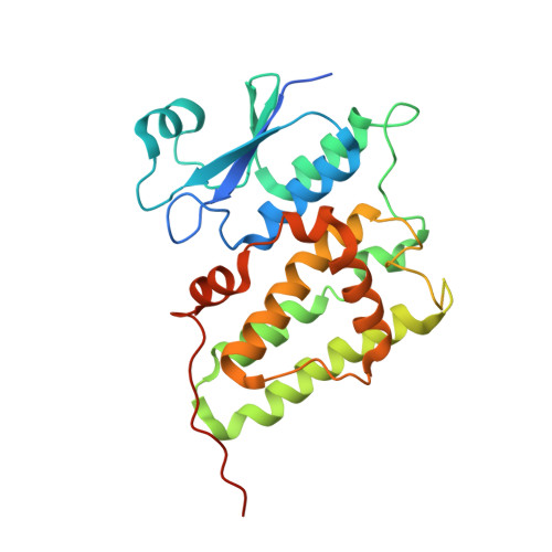
Crystal structure of the soluble form of the redox-regulated chloride ion channel protein CLIC4.

Littler, D.R.Assaad, N.N.Harrop, S.J.Brown, L.J.Pankhurst, G.J.Luciani, P.Aguilar, M.-I.Mazzanti, M.Berryman, M.A.Breit, S.N.Curmi, P.M.G.

(2005) FEBS J 272: 4996-5007

  • DOI: https://doi.org/10.1111/j.1742-4658.2005.04909.x
  • Primary Citation of Related Structures:  
    2AHE

  • PubMed Abstract: 

    The structure of CLIC4, a member of the CLIC family of putative intracellular chloride ion channel proteins, has been determined at 1.8 Angstroms resolution by X-ray crystallography. The protein is monomeric and it is structurally similar to CLIC1, belonging to the GST fold class. Differences between the structures of CLIC1 and CLIC4 are localized to helix 2 in the glutaredoxin-like N-terminal domain, which has previously been shown to undergo a dramatic structural change in CLIC1 upon oxidation. The structural differences in this region correlate with the sequence differences, where the CLIC1 sequence appears to be atypical of the family. Purified, recombinant, wild-type CLIC4 is shown to bind to artificial lipid bilayers, induce a chloride efflux current when associated with artificial liposomes and produce an ion channel in artificial bilayers with a conductance of 30 pS. Membrane binding is enhanced by oxidation of CLIC4 while no channels were observed via tip-dip electrophysiology in the presence of a reducing agent. Thus, recombinant CLIC4 appears to be able to form a redox-regulated ion channel in the absence of any partner proteins.


  • Organizational Affiliation

    School of Physics, University of New South Wales, Sydney, Australia.


Macromolecules
Find similar proteins by:  (by identity cutoff)  |  3D Structure
Entity ID: 1
MoleculeChains Sequence LengthOrganismDetailsImage
Chloride intracellular channel protein 4267Homo sapiensMutation(s): 0 
UniProt & NIH Common Fund Data Resources
Find proteins for Q9Y696 (Homo sapiens)
Explore Q9Y696 
Go to UniProtKB:  Q9Y696
PHAROS:  Q9Y696
GTEx:  ENSG00000169504 
Entity Groups  
Sequence Clusters30% Identity50% Identity70% Identity90% Identity95% Identity100% Identity
UniProt GroupQ9Y696
Sequence Annotations
Expand
  • Reference Sequence
Experimental Data & Validation

Experimental Data

  • Method: X-RAY DIFFRACTION
  • Resolution: 1.80 Å
  • R-Value Free: 0.229 
  • R-Value Work: 0.194 
  • R-Value Observed: 0.196 
  • Space Group: P 21 21 2
Unit Cell:
Length ( Å )Angle ( ˚ )
a = 77.717α = 90
b = 79.478β = 90
c = 42.6γ = 90
Software Package:
Software NamePurpose
REFMACrefinement
MOSFLMdata reduction
CCP4data scaling
AMoREphasing

Structure Validation

View Full Validation Report


Entry History 

Revision History  (Full details and data files)

  • Version 1.0: 2005-08-16
    Type: Initial release
  • Version 1.1: 2008-04-30
    Changes: Version format compliance
  • Version 1.2: 2011-07-13
    Changes: Version format compliance
  • Version 1.3: 2023-10-25
    Changes: Data collection, Database references, Refinement description