


2AEC
Crystal Structure of Human M340H-Beta1,4-Galactosyltransferase-I (M340H-B4GAL-T1) in Complex with GlcNAc-beta1,2-Man-alpha1,6-Man-beta-OR
- PDB DOI: https://doi.org/10.2210/pdb2AEC/pdb
- Classification: TRANSFERASE
- Organism(s): Homo sapiens
- Expression System: Escherichia coli BL21
- Mutation(s): Yes
- Deposited: 2005-07-22 Released: 2005-10-04
Experimental Data Snapshot
- Method: X-RAY DIFFRACTION
- Resolution: 2.00 Å
- R-Value Free: 0.239
- R-Value Work: 0.208
- R-Value Observed: 0.208

This is version 2.2 of the entry. See complete history.

Macromolecules
Find similar proteins by:
(by identity cutoff) | 3D Structure
Entity ID: 1 | |||||
|---|---|---|---|---|---|
| Molecule | Chains | Sequence Length | Organism | Details | Image |
| Beta-1,4-galactosyltransferase 1 | 287 | Homo sapiens | Mutation(s): 3 Gene Names: B4GALT1, GGTB2 EC: 2.4.1.90 | 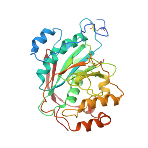 | |
UniProt & NIH Common Fund Data Resources | |||||
Find proteins for P15291 (Homo sapiens) Explore P15291 Go to UniProtKB: P15291 | |||||
PHAROS: P15291 GTEx: ENSG00000086062 | |||||
Entity Groups | |||||
| Sequence Clusters | 30% Identity50% Identity70% Identity90% Identity95% Identity100% Identity | ||||
| UniProt Group | P15291 | ||||
Sequence AnnotationsExpand | |||||
| |||||
Oligosaccharides

Small Molecules
| Ligands 6 Unique | |||||
|---|---|---|---|---|---|
| ID | Chains | Name / Formula / InChI Key | 2D Diagram | 3D Interactions | |
| UDH Query on UDH | FA [auth C], L [auth A], V [auth B] | 6-AMINOHEXYL-URIDINE-C1,5'-DIPHOSPHATE C15 H27 N3 O12 P2 MLWJBKPFDKRHBM-FMKGYKFTSA-N |  | ||
| MES Query on MES | HA [auth C] | 2-(N-MORPHOLINO)-ETHANESULFONIC ACID C6 H13 N O4 S SXGZJKUKBWWHRA-UHFFFAOYSA-N |  | ||
| SO4 Query on SO4 | BA [auth C] CA [auth C] DA [auth C] EA [auth C] H [auth A] | SULFATE ION O4 S QAOWNCQODCNURD-UHFFFAOYSA-L |  | ||
| GOL Query on GOL | IA [auth C] M [auth A] N [auth A] W [auth B] X [auth B] | GLYCEROL C3 H8 O3 PEDCQBHIVMGVHV-UHFFFAOYSA-N |  | ||
| DIO Query on DIO | GA [auth C] | 1,4-DIETHYLENE DIOXIDE C4 H8 O2 RYHBNJHYFVUHQT-UHFFFAOYSA-N |  | ||
| MN Query on MN | AA [auth C], G [auth A], O [auth B] | MANGANESE (II) ION Mn WAEMQWOKJMHJLA-UHFFFAOYSA-N |  | ||
Experimental Data & Validation
Experimental Data
- Method: X-RAY DIFFRACTION
- Resolution: 2.00 Å
- R-Value Free: 0.239
- R-Value Work: 0.208
- R-Value Observed: 0.208
- Space Group: C 2 2 21
Unit Cell:
| Length ( Å ) | Angle ( ˚ ) |
|---|---|
| a = 107.228 | α = 90 |
| b = 194.99 | β = 90 |
| c = 143.744 | γ = 90 |
| Software Name | Purpose |
|---|---|
| HKL-2000 | data collection |
| SCALEPACK | data scaling |
| AMoRE | phasing |
| CNS | refinement |
| HKL-2000 | data reduction |
Entry History
Deposition Data
- Released Date: 2005-10-04 Deposition Author(s): Ramasamy, V., Ramakrishnan, B., Boeggeman, E., Ratner, D.M., Seeberger, P.H., Qasba, P.K.
Revision History (Full details and data files)
- Version 1.0: 2005-10-04
Type: Initial release - Version 1.1: 2008-04-30
Changes: Version format compliance - Version 1.2: 2011-07-13
Changes: Non-polymer description, Version format compliance - Version 2.0: 2020-07-29
Type: Remediation
Reason: Carbohydrate remediation
Changes: Atomic model, Data collection, Derived calculations, Structure summary - Version 2.1: 2021-10-20
Changes: Database references, Structure summary - Version 2.2: 2023-08-23
Changes: Data collection, Refinement description
