Tyrosine-48 is the proton donor and histidine-110 directs substrate stereochemical selectivity in the reduction reaction of human aldose reductase: enzyme kinetics and crystal structure of the Y48H mutant enzyme.
Bohren, K.M., Grimshaw, C.E., Lai, C.J., Harrison, D.H., Ringe, D., Petsko, G.A., Gabbay, K.H.(1994) Biochemistry 33: 2021-2032
- PubMed: 8117659
- DOI: https://doi.org/10.1021/bi00174a007
- Primary Citation of Related Structures:
2ACU - PubMed Abstract:
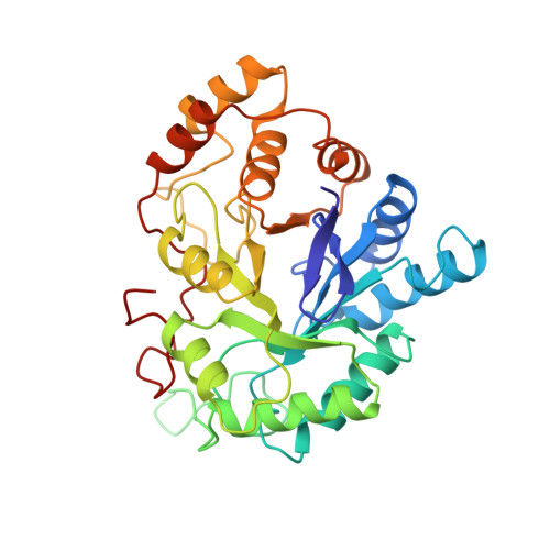
The active site of human aldose reductase contains two residues, His110 and Tyr48, either of which could be the proton donor during catalysis. Tyr48 is a candidate since its hydroxyl group is in proximity to Lys77 and thus may have an abnormally low pKa value. To distinguish between these possibilities, we used site-directed mutagenesis to create the H110Q and H110A, the Y48F, Y48H, and Y48S, and the K77M mutant enzymes. The two His110 mutants resulted in a 1000-20,000-fold drop in kcat/Km, respectively, for the reduction of DL-glyceraldehyde at pH 7. The Y48F mutation caused total loss of activity, whereas the Y48H and Y48S mutants retained catalytic activity with kcat/Km reduced by 5 orders of magnitude. The K77M mutant is an inactive enzyme. Kinetic studies using xylose stereoisomers show that the wild-type enzyme distinguishes between D-xylose, L-xylose, and D-lyxose up to 150-fold better than the H110A or H110Q mutants. The His110 mutants do not effectively discriminate between these isomers (4-11-fold). The crystal structure of the Y48H mutant refined at 1.8-A resolution shows that the overall structure is not significantly different from the wild-type structure. Electron densities for the histidine side chain and a new water molecule fill the space occupied by Tyr48 in the wild-type enzyme. The water molecule is in hydrogen-bonding distance to the N zeta group of Lys77 and to the N epsilon of His48 and fills the space occupied by the hydroxyl group of tyrosine in the wild-type structure. These findings suggest that proton transfer is mediated in the Y48H mutant enzyme by the water molecule. The Y48H mutant shows large and equal primary deuterium isotope effects on kcat and kcat/Km (1.81 +/- 0.03), providing direct evidence for hydride transfer as the rate-determining step in this mutant. Deuterium solvent isotope effects indicate that the relative contribution of proton transfer to this step of the catalytic cascade is much less important for the Y48H mutant than for the wild-type enzyme [D2O(kcat/Km) = 1.06 +/- 0.02 and 4.73 +/- 0.23, respectively]. The kinetic and mutagenesis data, together with structural data, indicate that His 110 plays an important role in the orientation of substrates in the active site pocket, while Tyr48 is the proton donor during aldehyde reduction by aldose reductase.
Organizational Affiliation:
Department of Pediatrics, Baylor College of Medicine, Houston, Texas 77030.