2ABS

Crystal structure of T. gondii adenosine kinase complexed with AMP-PCP


Experimental Data Snapshot

  • Method: X-RAY DIFFRACTION
  • Resolution: 1.10 Å
  • R-Value Free: 0.167 
  • R-Value Work: 0.135 
  • R-Value Observed: 0.137 

wwPDB Validation   3D Report Full Report


Ligand Structure Quality Assessment 


This is version 1.5 of the entry. See complete history


Literature

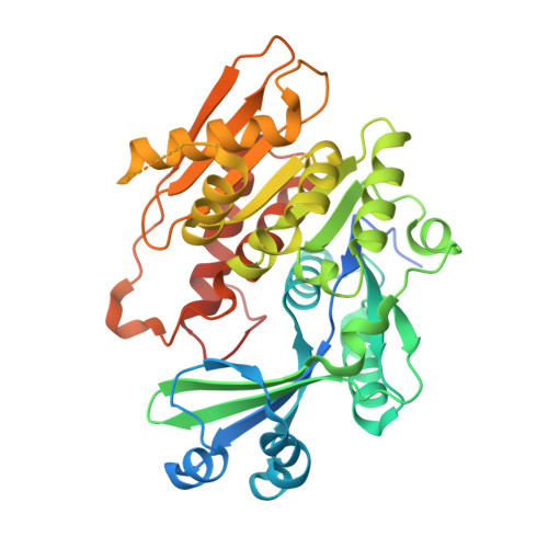
Structure of Toxoplasma gondii adenosine kinase in complex with an ATP analog at 1.1 angstroms resolution.

Zhang, Y.El Kouni, M.H.Ealick, S.E.

(2006) Acta Crystallogr D Biol Crystallogr 62: 140-145

  • DOI: https://doi.org/10.1107/S090744490503430X
  • Primary Citation of Related Structures:  
    2ABS

  • PubMed Abstract: 

    The obligate intracellular parasite Toxoplasma gondii is incapable of synthesizing purine nucleotides de novo and relies completely on purines salvaged from the host cells. Adenosine is the preferred precursor and is phosphorylated by adenosine kinase (AK), the most active enzyme in adenosine metabolism in T. gondii. AK thus represents a potential chemotherapeutic target for the treatment of T. gondii infections. The previously solved structures of unliganded AK and AK in complex with adenosine (or 7-iodotubercidin) and an ATP analog revealed a novel catalytic mechanism. A domain closure triggered by a GG switch upon adenosine binding sequesters the adenosine and gamma-phosphate of ATP from the solvent. The formation of the anion hole induced by the ATP binding completes the structural requirements for catalysis. In the current study, the structure of a binary complex of AK and the non-hydrolysable ATP analog AMP-PCP was determined to 1.1 angstroms resolution. The overall structure is similar to the apoenzyme, with an open conformation. AMP-PCP is bound in two relaxed conformations and without anchoring by Arg136. The induced anion hole is the same as that in the ternary complex AK-adenosine-AMP-PCP. This structure provides direct evidence that ATP binding at millimolar concentrations does not require adenosine binding as a prerequisite.


  • Organizational Affiliation

    Department of Molecular Biology and Genetics, Cornell University, Ithaca, NY 14853, USA.


Macromolecules
Find similar proteins by:  (by identity cutoff)  |  3D Structure
Entity ID: 1
MoleculeChains Sequence LengthOrganismDetailsImage
adenosine kinase383Toxoplasma gondiiMutation(s): 5 
Gene Names: AK
EC: 2.7.1.20
UniProt
Find proteins for Q9TVW2 (Toxoplasma gondii)
Explore Q9TVW2 
Go to UniProtKB:  Q9TVW2
Entity Groups  
Sequence Clusters30% Identity50% Identity70% Identity90% Identity95% Identity100% Identity
UniProt GroupQ9TVW2
Sequence Annotations
Expand
  • Reference Sequence
Experimental Data & Validation

Experimental Data

  • Method: X-RAY DIFFRACTION
  • Resolution: 1.10 Å
  • R-Value Free: 0.167 
  • R-Value Work: 0.135 
  • R-Value Observed: 0.137 
  • Space Group: P 21 21 21
Unit Cell:
Length ( Å )Angle ( ˚ )
a = 60.051α = 90
b = 67.923β = 90
c = 83.192γ = 90
Software Package:
Software NamePurpose
SHELXL-97refinement
SCALEPACKdata scaling
CNSrefinement
HKL-2000data collection
HKL-2000data reduction
CNSphasing

Structure Validation

View Full Validation Report


Ligand Structure Quality Assessment 


Entry History 

Deposition Data

Revision History  (Full details and data files)

  • Version 1.0: 2006-01-24
    Type: Initial release
  • Version 1.1: 2008-04-30
    Changes: Version format compliance
  • Version 1.2: 2011-07-13
    Changes: Version format compliance
  • Version 1.3: 2019-07-24
    Changes: Data collection, Refinement description
  • Version 1.4: 2021-10-20
    Changes: Database references, Derived calculations
  • Version 1.5: 2023-08-23
    Changes: Data collection, Refinement description