2A9Z

Crystal structure of T. gondii adenosine kinase complexed with N6-dimethyladenosine and AMP-PCP


Experimental Data Snapshot

  • Method: X-RAY DIFFRACTION
  • Resolution: 1.35 Å
  • R-Value Free: 0.196 
  • R-Value Work: 0.137 
  • R-Value Observed: 0.141 

wwPDB Validation   3D Report Full Report


Ligand Structure Quality Assessment 


This is version 1.5 of the entry. See complete history


Literature

Substrate analogs induce an intermediate conformational change in Toxoplasma gondii adenosine kinase

Zhang, Y.El Kouni, M.H.Ealick, S.E.

(2007) Acta Crystallogr D Biol Crystallogr 63: 126-134

  • DOI: https://doi.org/10.1107/S0907444906043654
  • Primary Citation of Related Structures:  
    2A9Y, 2A9Z, 2AA0, 2AB8

  • PubMed Abstract: 

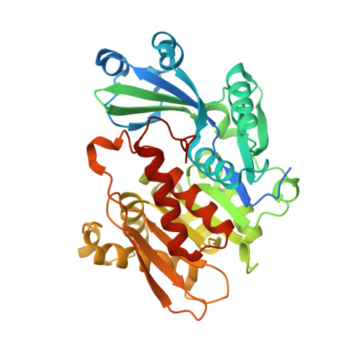
    Adenosine kinase (AK) is a key enzyme in purine metabolism in the ubiquitous intracellular parasite Toxoplasma gondii and is a potential chemotherapeutic target for the treatment of T. gondii infections. To better understand the structure-activity relationship of 6-substituted purine ribosides, the structures of the T. gondii AK-N6,N6-dimethyladenosine (DMA) complex, the AK-DMA-AMP-PCP complex, the AK-6-methyl mercaptopurine riboside (MMPR) complex and the AK-MMPR-AMP-PCP complex were determined to 1.35, 1.35, 1.75 and 1.75 A resolution, respectively. These structures reveal a conformation intermediate between open and closed, with a small lid-domain rotation of 12 degrees . Residues Gly143-X-X-Gly146 undergo torsional changes upon substrate binding, which together with a Gly68-Gly69 switch induces a hinge bending of the lid domain. The intermediate conformation suggests that ATP binding is independent of adenosine binding. Orienting the gamma-phosphate group of ATP into the optimal catalytic position may be the last step before the onset of chemical catalysis and may require the translocation of Arg136 following the complete closure of the lid domain. 6-Substituted purine-nucleoside analogs are accommodated in a hydrophobic cavity. Modification at the N6 or C6 position of the nucleoside would affect the interactions with the surrounding residues and the binding affinity.


  • Organizational Affiliation

    Department of Molecular Biology and Genetics, Cornell University, Ithaca, NY 14853, USA.


Macromolecules
Find similar proteins by:  (by identity cutoff)  |  3D Structure
Entity ID: 1
MoleculeChains Sequence LengthOrganismDetailsImage
adenosine kinase383Toxoplasma gondiiMutation(s): 5 
Gene Names: AK
EC: 2.7.1.20
UniProt
Find proteins for Q9TVW2 (Toxoplasma gondii)
Explore Q9TVW2 
Go to UniProtKB:  Q9TVW2
Entity Groups  
Sequence Clusters30% Identity50% Identity70% Identity90% Identity95% Identity100% Identity
UniProt GroupQ9TVW2
Sequence Annotations
Expand
  • Reference Sequence
Experimental Data & Validation

Experimental Data

  • Method: X-RAY DIFFRACTION
  • Resolution: 1.35 Å
  • R-Value Free: 0.196 
  • R-Value Work: 0.137 
  • R-Value Observed: 0.141 
  • Space Group: P 21 21 21
Unit Cell:
Length ( Å )Angle ( ˚ )
a = 60.068α = 90
b = 61.588β = 90
c = 91.65γ = 90
Software Package:
Software NamePurpose
SHELXL-97refinement
SCALEPACKdata scaling
CNSrefinement
HKL-2000data collection
HKL-2000data reduction
CNSphasing

Structure Validation

View Full Validation Report


Ligand Structure Quality Assessment 


Entry History 

Deposition Data

Revision History  (Full details and data files)

  • Version 1.0: 2006-07-11
    Type: Initial release
  • Version 1.1: 2008-04-30
    Changes: Version format compliance
  • Version 1.2: 2011-07-13
    Changes: Version format compliance
  • Version 1.3: 2019-07-24
    Changes: Data collection, Refinement description
  • Version 1.4: 2021-10-20
    Changes: Database references, Derived calculations
  • Version 1.5: 2023-08-23
    Changes: Data collection, Refinement description