2A5H

2.1 Angstrom X-ray crystal structure of lysine-2,3-aminomutase from Clostridium subterminale SB4, with Michaelis analog (L-alpha-lysine external aldimine form of pyridoxal-5'-phosphate).


Experimental Data Snapshot

  • Method: X-RAY DIFFRACTION
  • Resolution: 2.10 Å
  • R-Value Free: 0.225 
  • R-Value Work: 0.184 
  • R-Value Observed: 0.187 

wwPDB Validation   3D Report Full Report


Ligand Structure Quality Assessment 


This is version 1.3 of the entry. See complete history


Literature

The X-ray crystal structure of lysine-2,3-aminomutase from Clostridium subterminale.

Lepore, B.W.Ruzicka, F.J.Frey, P.A.Ringe, D.

(2005) Proc Natl Acad Sci U S A 102: 13819-13824

  • DOI: https://doi.org/10.1073/pnas.0505726102
  • Primary Citation of Related Structures:  
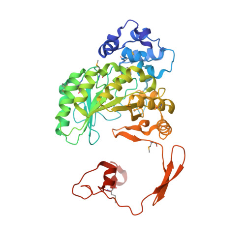
    2A5H

  • PubMed Abstract: 

    The x-ray crystal structure of the pyridoxal-5'-phosphate (PLP), S-adenosyl-L-methionine (SAM), and [4Fe-4S]-dependent lysine-2,3-aminomutase (LAM) of Clostridium subterminale has been solved to 2.1-A resolution by single-wavelength anomalous dispersion methods on a L-selenomethionine-substituted complex of LAM with [4Fe-4S]2+, PLP, SAM, and L-alpha-lysine, a very close analog of the active Michaelis complex. The unit cell contains a dimer of hydrogen-bonded, domain-swapped dimers, the subunits of which adopt a fold that contains all three cofactors in a central channel defined by six beta/alpha structural units. Zinc coordination links the domain-swapped dimers. In each subunit, the solvent face of the channel is occluded by an N-terminal helical domain, with the opposite end of the channel packed against the domain-swapped subunit. Hydrogen-bonded ionic contacts hold the external aldimine of PLP and L-alpha-lysine in position for abstraction of the 3-pro-R hydrogen of lysine by C5' of SAM. The structure of the SAM/[4Fe-4S] complex confirms and extends conclusions from spectroscopic studies of LAM and shows selenium in Se-adenosyl-L-selenomethionine poised to ligate the unique iron in the [4Fe-4S] cluster upon electron transfer and radical formation. The chain fold in the central domain is in part analogous to other radical-SAM enzymes.


  • Organizational Affiliation

    Department of Biochemistry, College of Agricultural and Life Sciences, University of Wisconsin, 1710 University Avenue, Madison, WI 53726, USA.


Macromolecules
Find similar proteins by:  (by identity cutoff)  |  3D Structure
Entity ID: 1
MoleculeChains Sequence LengthOrganismDetailsImage
L-lysine 2,3-aminomutase
A, B, C, D
416Clostridium subterminaleMutation(s): 10 
Gene Names: KamA
EC: 5.4.3.2
UniProt
Find proteins for Q9XBQ8 (Clostridium subterminale)
Explore Q9XBQ8 
Go to UniProtKB:  Q9XBQ8
Entity Groups  
Sequence Clusters30% Identity50% Identity70% Identity90% Identity95% Identity100% Identity
UniProt GroupQ9XBQ8
Sequence Annotations
Expand
  • Reference Sequence
Small Molecules
Ligands 6 Unique
IDChains Name / Formula / InChI Key2D Diagram3D Interactions
SAM
Query on SAM

Download Ideal Coordinates CCD File 
G [auth A],
M [auth B],
S [auth C],
Y [auth D]
S-ADENOSYLMETHIONINE
C15 H22 N6 O5 S
MEFKEPWMEQBLKI-FCKMPRQPSA-N
SF4
Query on SF4

Download Ideal Coordinates CCD File 
BA [auth D],
J [auth A],
P [auth B],
V [auth C]
IRON/SULFUR CLUSTER
Fe4 S4
LJBDFODJNLIPKO-UHFFFAOYSA-N
PLP
Query on PLP

Download Ideal Coordinates CCD File 
AA [auth D],
I [auth A],
O [auth B],
U [auth C]
PYRIDOXAL-5'-PHOSPHATE
C8 H10 N O6 P
NGVDGCNFYWLIFO-UHFFFAOYSA-N
LYS
Query on LYS

Download Ideal Coordinates CCD File 
H [auth A],
N [auth B],
T [auth C],
Z [auth D]
LYSINE
C6 H15 N2 O2
KDXKERNSBIXSRK-YFKPBYRVSA-O
SO4
Query on SO4

Download Ideal Coordinates CCD File 
F [auth A],
L [auth B],
Q [auth C],
W [auth D]
SULFATE ION
O4 S
QAOWNCQODCNURD-UHFFFAOYSA-L
ZN
Query on ZN

Download Ideal Coordinates CCD File 
E [auth A],
K [auth B],
R [auth C],
X [auth D]
ZINC ION
Zn
PTFCDOFLOPIGGS-UHFFFAOYSA-N
Modified Residues  1 Unique
IDChains TypeFormula2D DiagramParent
MSE
Query on MSE
A, B, C, D
L-PEPTIDE LINKINGC5 H11 N O2 SeMET
Experimental Data & Validation

Experimental Data

  • Method: X-RAY DIFFRACTION
  • Resolution: 2.10 Å
  • R-Value Free: 0.225 
  • R-Value Work: 0.184 
  • R-Value Observed: 0.187 
  • Space Group: C 1 2 1
Unit Cell:
Length ( Å )Angle ( ˚ )
a = 118.89α = 90
b = 92.926β = 96.74
c = 177.735γ = 90
Software Package:
Software NamePurpose
REFMACrefinement
CBASSdata collection
MXdata reduction
JBluIce-EPICSdata collection
SCALEPACKdata scaling
SOLVEphasing

Structure Validation

View Full Validation Report


Ligand Structure Quality Assessment 


Entry History 

Deposition Data

Revision History  (Full details and data files)

  • Version 1.0: 2005-10-04
    Type: Initial release
  • Version 1.1: 2008-04-30
    Changes: Version format compliance
  • Version 1.2: 2011-07-13
    Changes: Non-polymer description, Version format compliance
  • Version 1.3: 2017-10-11
    Changes: Advisory, Refinement description