2A4V

Crystal Structure of a truncated mutant of yeast nuclear thiol peroxidase


Experimental Data Snapshot

  • Method: X-RAY DIFFRACTION
  • Resolution: 1.80 Å
  • R-Value Free: 0.215 
  • R-Value Work: 0.165 
  • R-Value Observed: 0.167 

wwPDB Validation   3D Report Full Report


This is version 1.4 of the entry. See complete history


Literature

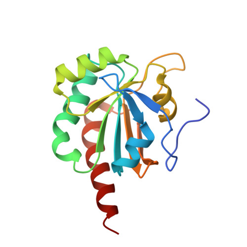
Crystal structure of the C107S/C112S mutant of yeast nuclear 2-Cys peroxiredoxin

Choi, J.Choi, S.Chon, J.-K.Choi, J.Cha, M.-K.Kim, I.-H.Shin, W.

(2005) Proteins 61: 1146-1149


Macromolecules
Find similar proteins by:  (by identity cutoff)  |  3D Structure
Entity ID: 1
MoleculeChains Sequence LengthOrganismDetailsImage
Peroxiredoxin DOT5159Saccharomyces cerevisiaeMutation(s): 3 
Gene Names: DOT5
EC: 1.11.1.15
UniProt
Find proteins for P40553 (Saccharomyces cerevisiae (strain ATCC 204508 / S288c))
Explore P40553 
Go to UniProtKB:  P40553
Entity Groups  
Sequence Clusters30% Identity50% Identity70% Identity90% Identity95% Identity100% Identity
UniProt GroupP40553
Sequence Annotations
Expand
  • Reference Sequence
Experimental Data & Validation

Experimental Data

  • Method: X-RAY DIFFRACTION
  • Resolution: 1.80 Å
  • R-Value Free: 0.215 
  • R-Value Work: 0.165 
  • R-Value Observed: 0.167 
  • Space Group: P 32
Unit Cell:
Length ( Å )Angle ( ˚ )
a = 37.535α = 90
b = 37.535β = 90
c = 83.256γ = 120
Software Package:
Software NamePurpose
DENZOdata reduction
SCALEPACKdata scaling
MOLREPphasing
REFMACrefinement
PDB_EXTRACTdata extraction
HKL-2000data reduction

Structure Validation

View Full Validation Report


Entry History 

Deposition Data

Revision History  (Full details and data files)

  • Version 1.0: 2006-03-14
    Type: Initial release
  • Version 1.1: 2008-04-30
    Changes: Version format compliance
  • Version 1.2: 2011-07-13
    Changes: Version format compliance
  • Version 1.3: 2017-10-11
    Changes: Refinement description
  • Version 1.4: 2021-11-10
    Changes: Database references