


Crystal Structure of Uridylate kinase
Gorman, J., Shapiro, L.To be published.
Experimental Data Snapshot
wwPDB Validation 3D Report Full Report

Entity ID: 1 | |||||
|---|---|---|---|---|---|
| Molecule | Chains | Sequence Length | Organism | Details | Image |
| Uridylate kinase | 247 | Haemophilus influenzae | Mutation(s): 0 Gene Names: pyrH, smbA EC: 2.7.4 | 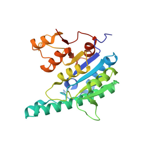 | |
UniProt | |||||
Find proteins for P43890 (Haemophilus influenzae (strain ATCC 51907 / DSM 11121 / KW20 / Rd)) Explore P43890 Go to UniProtKB: P43890 | |||||
Entity Groups | |||||
| Sequence Clusters | 30% Identity50% Identity70% Identity90% Identity95% Identity100% Identity | ||||
| UniProt Group | P43890 | ||||
Sequence AnnotationsExpand | |||||
| |||||
| Length ( Å ) | Angle ( ˚ ) |
|---|---|
| a = 77.369 | α = 94.85 |
| b = 79.889 | β = 96.68 |
| c = 79.899 | γ = 96.88 |
| Software Name | Purpose |
|---|---|
| REFMAC | refinement |
| DENZO | data reduction |
| SCALEPACK | data scaling |
| PHASER | phasing |
RCSB PDB (citation) is hosted by
RCSB PDB is a member of the

