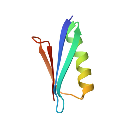
Optimizing pH response of affinity between protein G and IgG Fc: how electrostatic modulations affect protein-protein interactions.
Watanabe, H., Matsumaru, H., Ooishi, A., Feng, Y., Odahara, T., Suto, K., Honda, S.(2009) J Biol Chem 284: 12373-12383
- PubMed: 19269963
- DOI: https://doi.org/10.1074/jbc.M809236200
- Primary Citation of Related Structures:
2ZW0, 2ZW1 - PubMed Abstract:
Protein-protein interaction in response to environmental conditions enables sophisticated biological and biotechnological processes. Aiming toward the rational design of a pH-sensitive protein-protein interaction, we engineered pH-sensitive mutants of streptococcal protein G B1, a binder to the IgG constant region. We systematically introduced histidine residues into the binding interface to cause electrostatic repulsion on the basis of a rigid body model. Exquisite pH sensitivity of this interaction was confirmed by surface plasmon resonance and affinity chromatography employing a clinically used human IgG. The pH-sensitive mechanism of the interaction was analyzed and evaluated from kinetic, thermodynamic, and structural viewpoints. Histidine-mediated electrostatic repulsion resulted in significant loss of exothermic heat of the binding that decreased the affinity only at acidic conditions, thereby improving the pH sensitivity. The reduced binding energy was partly recovered by "enthalpy-entropy compensation." Crystal structures of the designed mutants confirmed the validity of the rigid body model on which the effective electrostatic repulsion was based. Moreover, our data suggested that the entropy gain involved exclusion of water molecules solvated in a space formed by the introduced histidine and adjacent tryptophan residue. Our findings concerning the mechanism of histidine-introduced interactions will provide a guideline for the rational design of pH-sensitive protein-protein recognition.
Organizational Affiliation:
National Institute of Advanced Industrial Science and Technology, Central 6, Tsukuba 305-8566, Japan.