2ZU0

Crystal structure of SufC-SufD complex involved in the iron-sulfur cluster biosynthesis


Experimental Data Snapshot

  • Method: X-RAY DIFFRACTION
  • Resolution: 2.20 Å
  • R-Value Free: 0.259 
  • R-Value Work: 0.234 
  • R-Value Observed: 0.234 

wwPDB Validation   3D Report Full Report


This is version 1.2 of the entry. See complete history


Literature

Molecular dynamism of Fe-S cluster biosynthesis implicated by the structure of SufC(2)-SufD(2) complex

Wada, K.Sumi, N.Nagai, R.Iwasaki, K.Sato, T.Suzuki, K.Hasegawa, Y.Kitaoka, S.Minami, Y.Outten, F.W.Takahashi, Y.Fukuyama, K.

(2009) J Mol Biol 387: 245-258

  • DOI: https://doi.org/10.1016/j.jmb.2009.01.054
  • Primary Citation of Related Structures:  
    2ZU0

  • PubMed Abstract: 

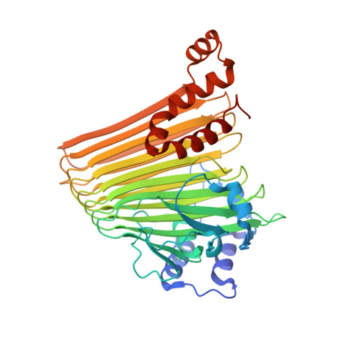
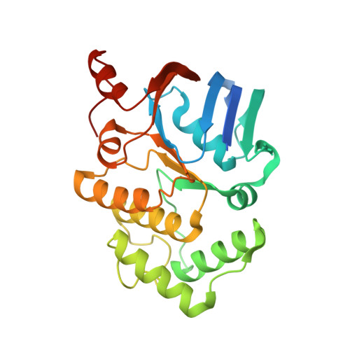
    Maturation of iron-sulfur (Fe-S) proteins is achieved by the SUF machinery in a wide number of eubacteria and archaea, as well as eukaryotic chloroplasts. This machinery is encoded in Escherichia coli by the sufABCDSE operon, where three Suf components, SufB, SufC, and SufD, form a complex and appear to provide an intermediary site for the Fe-S cluster assembly. Here, we report the quaternary structure of the SufC(2)-SufD(2) complex in which SufC is bound to the C-terminal domain of SufD. Comparison with the monomeric structure of SufC revealed conformational change of the active-site residues: SufC becomes competent for ATP binding and hydrolysis upon association with SufD. The two SufC subunits were spatially separated in the SufC(2)-SufD(2) complex, whereas cross-linking experiments in solution have indicated that two SufC molecules associate with each other in the presence of Mg(2+) and ATP. Such dimer formation of SufC may lead to a gross structural change of the SufC(2)-SufD(2) complex. Furthermore, genetic analysis of SufD revealed an essential histidine residue buried inside the dimer interface, suggesting that conformational change may expose this crucial residue. These findings, together with biochemical characterization of the SufB-SufC-SufD complex, have led us to propose a model for the Fe-S cluster biosynthesis in the complex.


  • Organizational Affiliation

    Department of Biological Sciences, Graduate School of Science, Osaka University, Toyonaka, Osaka, Japan.


Macromolecules
Find similar proteins by:  (by identity cutoff)  |  3D Structure
Entity ID: 1
MoleculeChains Sequence LengthOrganismDetailsImage
Protein sufD
A, B
423Escherichia coli K-12Mutation(s): 0 
Gene Names: SufC and SufD
UniProt
Find proteins for P77689 (Escherichia coli (strain K12))
Explore P77689 
Go to UniProtKB:  P77689
Entity Groups  
Sequence Clusters30% Identity50% Identity70% Identity90% Identity95% Identity100% Identity
UniProt GroupP77689
Sequence Annotations
Expand
  • Reference Sequence
Find similar proteins by:  (by identity cutoff)  |  3D Structure
Entity ID: 2
MoleculeChains Sequence LengthOrganismDetailsImage
Probable ATP-dependent transporter sufC
C, D
267Escherichia coli K-12Mutation(s): 0 
Gene Names: SufC and SufD
UniProt
Find proteins for P77499 (Escherichia coli (strain K12))
Explore P77499 
Go to UniProtKB:  P77499
Entity Groups  
Sequence Clusters30% Identity50% Identity70% Identity90% Identity95% Identity100% Identity
UniProt GroupP77499
Sequence Annotations
Expand
  • Reference Sequence
Small Molecules
Ligands 1 Unique
IDChains Name / Formula / InChI Key2D Diagram3D Interactions
MES
Query on MES

Download Ideal Coordinates CCD File 
E [auth C]2-(N-MORPHOLINO)-ETHANESULFONIC ACID
C6 H13 N O4 S
SXGZJKUKBWWHRA-UHFFFAOYSA-N
Experimental Data & Validation

Experimental Data

  • Method: X-RAY DIFFRACTION
  • Resolution: 2.20 Å
  • R-Value Free: 0.259 
  • R-Value Work: 0.234 
  • R-Value Observed: 0.234 
  • Space Group: P 21 21 21
Unit Cell:
Length ( Å )Angle ( ˚ )
a = 96.11α = 90
b = 106.15β = 90
c = 171.67γ = 90
Software Package:
Software NamePurpose
CNSrefinement
BSSdata collection
HKL-2000data reduction
HKL-2000data scaling
MOLREPphasing

Structure Validation

View Full Validation Report


Entry History 

Deposition Data

  • Released Date: 2009-03-10 
  • Deposition Author(s): Wada, K.

Revision History  (Full details and data files)

  • Version 1.0: 2009-03-10
    Type: Initial release
  • Version 1.1: 2011-07-13
    Changes: Version format compliance
  • Version 1.2: 2023-11-01
    Changes: Data collection, Database references, Derived calculations, Refinement description