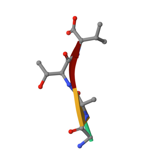
2Z9I

Crystal structure of RV0983 from Mycobacterium tuberculosis- Proteolytically active form


Experimental Data Snapshot

  • Method: X-RAY DIFFRACTION
  • Resolution: 2.00 Å
  • R-Value Free: 0.273 
  • R-Value Work: 0.225 
  • R-Value Observed: 0.228 

wwPDB Validation   3D Report Full Report


This is version 1.1 of the entry. See complete history


Literature

Structure and function of the virulence-associated high-temperature requirement A of Mycobacterium tuberculosis

Mohamedmohaideen, N.N.Palaninathan, S.K.Morin, P.M.Williams, B.J.Braunstein, M.Tichy, S.E.Locker, J.Russell, D.H.Jacobs, W.R.Sacchettini, J.C.

(2008) Biochemistry 47: 6092-6102

  • DOI: https://doi.org/10.1021/bi701929m
  • Primary Citation of Related Structures:  
    2Z9I

  • PubMed Abstract: 

    The high-temperature requirement A (HtrA) family of serine proteases has been shown to play an important role in the environmental and cellular stress damage control system in Escherichia coli. Mycobacterium tuberculosis ( Mtb) has three putative HtrA-like proteases, HtrA1, HtrA2, and HtrA3. The deletion of htrA2 gives attenuated virulence in a mouse model of TB. Biochemical analysis reveals that HtrA2 can function both as a protease and as a chaperone. The three-dimensional structure of HtrA2 determined at 2.0 A resolution shows that the protease domains form the central core of the trimer and the PDZ domains extend to the periphery. Unlike E. coli DegS and DegP, the protease is naturally active due to the formation of the serine protease-like catalytic triad and its uniquely designed oxyanion hole. Both protease and PDZ binding pockets of each HtrA2 molecule are occupied by autoproteolytic peptide products and reveal clues for a novel autoregulatory mechanism that might have significant importance in HtrA-associated virulence of Mtb.


  • Organizational Affiliation

    Department of Biochemistry and Biophysics, Texas A&M University, College Station, Texas 77843, USA.


Macromolecules
Find similar proteins by:  (by identity cutoff)  |  3D Structure
Entity ID: 1
MoleculeChains Sequence LengthOrganismDetailsImage
PROBABLE SERINE PROTEASE PEPD
A, B, C
324Mycobacterium tuberculosisMutation(s): 0 
EC: 3.4.21
UniProt
Find proteins for O53896 (Mycobacterium tuberculosis (strain ATCC 25618 / H37Rv))
Explore O53896 
Go to UniProtKB:  O53896
Entity Groups  
Sequence Clusters30% Identity50% Identity70% Identity90% Identity95% Identity100% Identity
UniProt GroupO53896
Sequence Annotations
Expand
  • Reference Sequence

Find similar proteins by:  Sequence   |   3D Structure  

Entity ID: 2
MoleculeChains Sequence LengthOrganismDetailsImage
SVEQV
D, E, F
5Mycobacterium tuberculosisMutation(s): 0 
Sequence Annotations
Expand
  • Reference Sequence

Find similar proteins by:  Sequence   |   3D Structure  

Entity ID: 3
MoleculeChains Sequence LengthOrganismDetailsImage
GATV
G, H, I
4Mycobacterium tuberculosisMutation(s): 0 
Sequence Annotations
Expand
  • Reference Sequence
Small Molecules
Modified Residues  1 Unique
IDChains TypeFormula2D DiagramParent
MSE
Query on MSE
A, B, C
L-PEPTIDE LINKINGC5 H11 N O2 SeMET
Experimental Data & Validation

Experimental Data

  • Method: X-RAY DIFFRACTION
  • Resolution: 2.00 Å
  • R-Value Free: 0.273 
  • R-Value Work: 0.225 
  • R-Value Observed: 0.228 
  • Space Group: C 1 2 1
Unit Cell:
Length ( Å )Angle ( ˚ )
a = 149.584α = 90
b = 89.067β = 97.55
c = 69.408γ = 90
Software Package:
Software NamePurpose
SHARPphasing
REFMACrefinement
HKL-2000data collection
HKL-2000data reduction
SCALEPACKdata scaling

Structure Validation

View Full Validation Report


Entry History 

Deposition Data

Revision History  (Full details and data files)

  • Version 1.0: 2008-06-10
    Type: Initial release
  • Version 1.1: 2011-07-13
    Changes: Version format compliance