2Z9A

Crystal Structure of Human Saposin C Dimer in Open Conformation


Experimental Data Snapshot

  • Method: X-RAY DIFFRACTION
  • Resolution: 2.50 Å
  • R-Value Free: 0.271 
  • R-Value Work: 0.239 
  • R-Value Observed: 0.240 

wwPDB Validation   3D Report Full Report


This is version 1.2 of the entry. See complete history


Literature

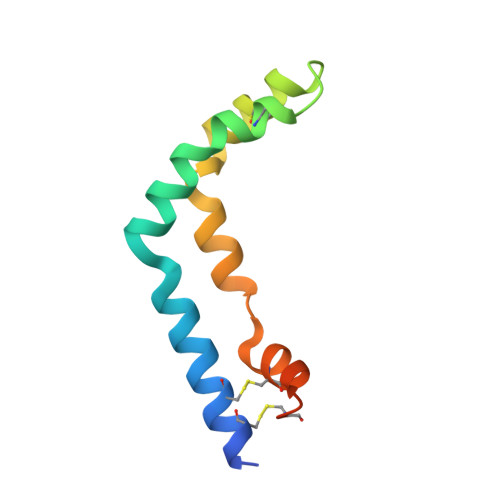
Crystal structures of human saposins C and d: implications for lipid recognition and membrane interactions.

Rossmann, M.Schultz-Heienbrok, R.Behlke, J.Remmel, N.Alings, C.Sandhoff, K.Saenger, W.Maier, T.

(2008) Structure 16: 809-817

  • DOI: https://doi.org/10.1016/j.str.2008.02.016
  • Primary Citation of Related Structures:  
    2QYP, 2R0R, 2R1Q, 2RB3, 2Z9A

  • PubMed Abstract: 

    Human saposins are essential proteins required for degradation of sphingolipids and lipid antigen presentation. Despite the conserved structural organization of saposins, their distinct modes of interaction with biological membranes are not fully understood. We describe two crystal structures of human saposin C in an "open" configuration with unusual domain swapped homodimers. This form of SapC dimer supports the "clip-on" model for SapC-induced vesicle fusion. In addition, we present the crystal structure of SapD in two crystal forms. They reveal the monomer-monomer interface for the SapD dimer, which was confirmed in solution by analytical ultracentrifugation. The crystal structure of SapD suggests that side chains of Lys10 and Arg17 are involved in initial association with the preferred anionic biological membranes by forming salt bridges with sulfate or phosphate lipid headgroups.


  • Organizational Affiliation

    Institut für Chemie und Biochemie, Kristallographie, Freie Universität Berlin, Takustr. 6, 14195 Berlin, Germany.


Macromolecules
Find similar proteins by:  (by identity cutoff)  |  3D Structure
Entity ID: 1
MoleculeChains Sequence LengthOrganismDetailsImage
Proactivator polypeptide
A, B
88Homo sapiensMutation(s): 0 
Gene Names: PSAPGLBASAP1
UniProt & NIH Common Fund Data Resources
Find proteins for P07602 (Homo sapiens)
Explore P07602 
Go to UniProtKB:  P07602
PHAROS:  P07602
GTEx:  ENSG00000197746 
Entity Groups  
Sequence Clusters30% Identity50% Identity70% Identity90% Identity95% Identity100% Identity
UniProt GroupP07602
Sequence Annotations
Expand
  • Reference Sequence
Experimental Data & Validation

Experimental Data

  • Method: X-RAY DIFFRACTION
  • Resolution: 2.50 Å
  • R-Value Free: 0.271 
  • R-Value Work: 0.239 
  • R-Value Observed: 0.240 
  • Space Group: P 41 21 2
Unit Cell:
Length ( Å )Angle ( ˚ )
a = 48.99α = 90
b = 48.99β = 90
c = 155.57γ = 90
Software Package:
Software NamePurpose
XSCALEdata scaling
REFMACrefinement
PDB_EXTRACTdata extraction
ADSCdata collection
XDSdata reduction
MOLREPphasing

Structure Validation

View Full Validation Report


Entry History 

Deposition Data

Revision History  (Full details and data files)

  • Version 1.0: 2008-04-29
    Type: Initial release
  • Version 1.1: 2011-07-13
    Changes: Non-polymer description, Version format compliance
  • Version 1.2: 2023-08-30
    Changes: Data collection, Database references, Derived calculations, Refinement description