2YKF

Sensor region of a sensor histidine kinase


Experimental Data Snapshot

  • Method: X-RAY DIFFRACTION
  • Resolution: 2.00 Å
  • R-Value Free: 0.240 
  • R-Value Work: 0.188 
  • R-Value Observed: 0.190 

wwPDB Validation   3D Report Full Report


This is version 1.3 of the entry. See complete history


Literature

The Sensor Region of the Ubiquitous Cytosolic Sensor Kinase, Pdtas, Contains Pas and Gaf Domain Sensing Modules.

Preu, J.Panjikar, S.Morth, P.Jaiswal, R.Karunakar, P.Tucker, P.A.

(2012) J Struct Biol 177: 498

  • DOI: https://doi.org/10.1016/j.jsb.2011.11.012
  • Primary Citation of Related Structures:  
    2YKF, 2YKH

  • PubMed Abstract: 

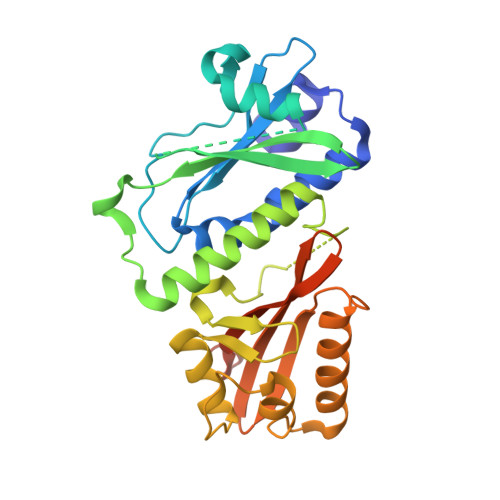
    Two-component systems, a sensor histidine kinase (HK) and a response regulator (RR), are ubiquitous signaling systems that allow prokaryotes to respond to external challenges. HKs normally have sensing modules and highly conserved cytosolic histidine kinase and ATPase domains. The interaction between the activated phosphohistidine and the cognate RR allows an external signal to be passed from the exterior of gram-positive bacteria (GPB) to the cytoplasm. Orthologs of the PdtaS/PdtaR regulatory system, found in most GPB phyla, are unusual in two respects. The HK is not membrane anchored, and the RR acts at the level of transcriptional antitermination. The structure of the complete sensor region of the cytosolic HK, PdtaS, from Mycobacterium tuberculosis consists of closely linked GAF and PAS domains. The structure and sequence analysis suggest that the PdtaS/PdtaR regulatory system is structurally equivalent to the EutW/EutV system regulating ethanolamine catabolism in some phyla but that the effector for the PAS domain is not ethanolamine in the Actinobacteria.


  • Organizational Affiliation

    EMBL Hamburg Outstation, c/o DESY, Notkestrasse 85, D22603 Hamburg, Germany.


Macromolecules
Find similar proteins by:  (by identity cutoff)  |  3D Structure
Entity ID: 1
MoleculeChains Sequence LengthOrganismDetailsImage
PROBABLE SENSOR HISTIDINE KINASE PDTAS305Mycobacterium tuberculosis H37RvMutation(s): 0 
EC: 2.7.13.3
UniProt
Find proteins for P9WGL5 (Mycobacterium tuberculosis (strain ATCC 25618 / H37Rv))
Explore P9WGL5 
Go to UniProtKB:  P9WGL5
Entity Groups  
Sequence Clusters30% Identity50% Identity70% Identity90% Identity95% Identity100% Identity
UniProt GroupP9WGL5
Sequence Annotations
Expand
  • Reference Sequence
Small Molecules
Ligands 2 Unique
IDChains Name / Formula / InChI Key2D Diagram3D Interactions
SO4
Query on SO4

Download Ideal Coordinates CCD File 
B [auth A]SULFATE ION
O4 S
QAOWNCQODCNURD-UHFFFAOYSA-L
BR
Query on BR

Download Ideal Coordinates CCD File 
C [auth A]
D [auth A]
E [auth A]
F [auth A]
G [auth A]
C [auth A],
D [auth A],
E [auth A],
F [auth A],
G [auth A],
H [auth A],
I [auth A],
J [auth A],
K [auth A],
L [auth A],
M [auth A],
N [auth A],
O [auth A],
P [auth A],
Q [auth A],
R [auth A]
BROMIDE ION
Br
CPELXLSAUQHCOX-UHFFFAOYSA-M
Experimental Data & Validation

Experimental Data

  • Method: X-RAY DIFFRACTION
  • Resolution: 2.00 Å
  • R-Value Free: 0.240 
  • R-Value Work: 0.188 
  • R-Value Observed: 0.190 
  • Space Group: C 2 2 21
Unit Cell:
Length ( Å )Angle ( ˚ )
a = 73.59α = 90
b = 81.27β = 90
c = 105.06γ = 90
Software Package:
Software NamePurpose
REFMACrefinement
DENZOdata reduction
XDSdata reduction
SCALEPACKdata scaling
XSCALEdata scaling
Auto-Rickshawphasing

Structure Validation

View Full Validation Report


Entry History 

Deposition Data

Revision History  (Full details and data files)

  • Version 1.0: 2012-06-06
    Type: Initial release
  • Version 1.1: 2012-06-27
    Changes: Other
  • Version 1.2: 2015-09-23
    Changes: Data collection, Other
  • Version 1.3: 2019-07-17
    Changes: Data collection