2YDJ

Discovery of Checkpoint Kinase Inhibitor AZD7762 by Structure Based Design and Optimization of Thiophene Carboxamide Ureas


Experimental Data Snapshot

  • Method: X-RAY DIFFRACTION
  • Resolution: 1.85 Å
  • R-Value Free: 0.274 
  • R-Value Work: 0.229 
  • R-Value Observed: 0.231 

wwPDB Validation   3D Report Full Report


Ligand Structure Quality Assessment 


This is version 1.4 of the entry. See complete history


Literature

Discovery of Checkpoint Kinase Inhibitor (S)-5-(3-Fluorophenyl)-N-(Piperidin-3-Yl)-3-Ureidothiophene-2-Carboxamide (Azd7762) by Structure-Based Design and Optimization of Thiophenecarboxamide Ureas.

Oza, V.Ashwell, S.Almeida, L.Brassil, P.Breed, J.Deng, C.Gero, T.Grondine, M.Horn, C.Ioannidis, S.Liu, D.Lyne, P.Newcombe, N.Pass, M.Read, J.A.Ready, S.Rowsell, S.Su, M.Toader, D.Vasbinder, M.Yu, D.Yu, Y.Xue, Y.Zabludoff, S.Janetka, J.

(2012) J Med Chem 55: 5130

  • DOI: https://doi.org/10.1021/jm300025r
  • Primary Citation of Related Structures:  
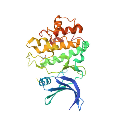
    2YDI, 2YDJ, 2YDK, 3PZE

  • PubMed Abstract: 

    Checkpoint kinases CHK1 and CHK2 are activated in response to DNA damage that results in cell cycle arrest, allowing sufficient time for DNA repair. Agents that lead to abrogation of such checkpoints have potential to increase the efficacy of such compounds as chemo- and radiotherapies. Thiophenecarboxamide ureas (TCUs) were identified as inhibitors of CHK1 by high throughput screening. A structure-based approach is described using crystal structures of JNK1 and CHK1 in complex with 1 and 2 and of the CHK1-3b complex. The ribose binding pocket of CHK1 was targeted to generate inhibitors with excellent cellular potency and selectivity over CDK1and IKKβ, key features lacking from the initial compounds. Optimization of 3b resulted in the identification of a regioisomeric 3-TCU lead 12a. Optimization of 12a led to the discovery of the clinical candidate 4 (AZD7762), which strongly potentiates the efficacy of a variety of DNA-damaging agents in preclinical models.


  • Organizational Affiliation

    AstraZeneca R&D Boston, 35 Gatehouse Drive, Waltham, Massachusetts 02451, United States. vibha.oza@gmail.com


Macromolecules
Find similar proteins by:  (by identity cutoff)  |  3D Structure
Entity ID: 1
MoleculeChains Sequence LengthOrganismDetailsImage
SERINE/THREONINE-PROTEIN KINASE CHK1
A, B
276Homo sapiensMutation(s): 0 
EC: 2.7.11.1
UniProt & NIH Common Fund Data Resources
Find proteins for O14757 (Homo sapiens)
Explore O14757 
Go to UniProtKB:  O14757
PHAROS:  O14757
GTEx:  ENSG00000149554 
Entity Groups  
Sequence Clusters30% Identity50% Identity70% Identity90% Identity95% Identity100% Identity
UniProt GroupO14757
Sequence Annotations
Expand
  • Reference Sequence
Small Molecules
Modified Residues  1 Unique
IDChains TypeFormula2D DiagramParent
CSS
Query on CSS
A, B
L-PEPTIDE LINKINGC3 H7 N O2 S2CYS
Binding Affinity Annotations 
IDSourceBinding Affinity
YDJ PDBBind:  2YDJ IC50: 5 (nM) from 1 assay(s)
BindingDB:  2YDJ IC50: min: 5, max: 7.9 (nM) from 3 assay(s)
EC50: min: 10, max: 280 (nM) from 2 assay(s)
Binding MOAD:  2YDJ IC50: 5 (nM) from 1 assay(s)
Experimental Data & Validation

Experimental Data

  • Method: X-RAY DIFFRACTION
  • Resolution: 1.85 Å
  • R-Value Free: 0.274 
  • R-Value Work: 0.229 
  • R-Value Observed: 0.231 
  • Space Group: P 1 21 1
Unit Cell:
Length ( Å )Angle ( ˚ )
a = 42.004α = 90
b = 64.854β = 98.77
c = 119.999γ = 90
Software Package:
Software NamePurpose
REFMACrefinement
d*TREKdata reduction
d*TREKdata scaling
AMoREphasing

Structure Validation

View Full Validation Report


Ligand Structure Quality Assessment 


Entry History 

Deposition Data

Revision History  (Full details and data files)

  • Version 1.0: 2012-01-25
    Type: Initial release
  • Version 1.1: 2012-06-27
    Changes: Other
  • Version 1.2: 2013-03-27
    Changes: Non-polymer description
  • Version 1.3: 2017-06-28
    Changes: Data collection
  • Version 1.4: 2019-04-03
    Changes: Data collection, Derived calculations, Source and taxonomy