2YAY

The Crystal Structure of Leishmania major dUTPase in complex with substrate analogue dUpNpp


Experimental Data Snapshot

  • Method: X-RAY DIFFRACTION
  • Resolution: 1.86 Å
  • R-Value Free: 0.249 
  • R-Value Work: 0.214 
  • R-Value Observed: 0.216 

wwPDB Validation   3D Report Full Report


Ligand Structure Quality Assessment 


This is version 1.3 of the entry. See complete history


Literature

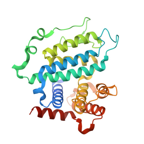
The Crystal Structure of the Leishmania Major Deoxyuridine Triphosphate Nucleotidohydrolase in Complex with Nucleotide Analogues, Dump, and Deoxyuridine.

Hemsworth, G.R.Moroz, O.V.Fogg, M.J.Scott, B.Bosch-Navarrete, C.Gonzalez-Pacanowska, D.Wilson, K.S.

(2011) J Biol Chem 286: 16470

  • DOI: https://doi.org/10.1074/jbc.M111.224873
  • Primary Citation of Related Structures:  
    2CIC, 2CJE, 2YAY, 2YAZ, 2YB0

  • PubMed Abstract: 

    Members of the Leishmania genus are the causative agents of the life-threatening disease leishmaniasis. New drugs are being sought due to increasing resistance and adverse side effects with current treatments. The knowledge that dUTPase is an essential enzyme and that the all α-helical dimeric kinetoplastid dUTPases have completely different structures compared with the trimeric β-sheet type dUTPase possessed by most organisms, including humans, make the dimeric enzymes attractive drug targets. Here, we present crystal structures of the Leishmania major dUTPase in complex with substrate analogues, the product dUMP and a substrate fragment, and of the homologous Campylobacter jejuni dUTPase in complex with a triphosphate substrate analogue. The metal-binding properties of both enzymes are shown to be dependent upon the ligand identity, a previously unseen characteristic of this family. Furthermore, structures of the Leishmania enzyme in the presence of dUMP and deoxyuridine coupled with tryptophan fluorescence quenching indicate that occupation of the phosphate binding region is essential for induction of the closed conformation and hence for substrate binding. These findings will aid in the development of dUTPase inhibitors as potential new lead anti-trypanosomal compounds.


  • Organizational Affiliation

    Structural Biology Laboratory, Department of Chemistry, University of York, Heslington, York, United Kingdom.


Macromolecules
Find similar proteins by:  (by identity cutoff)  |  3D Structure
Entity ID: 1
MoleculeChains Sequence LengthOrganismDetailsImage
DUTPASE271Leishmania majorMutation(s): 0 
EC: 3.6.1.23
UniProt
Find proteins for O15826 (Leishmania major)
Explore O15826 
Go to UniProtKB:  O15826
Entity Groups  
Sequence Clusters30% Identity50% Identity70% Identity90% Identity95% Identity100% Identity
UniProt GroupO15826
Sequence Annotations
Expand
  • Reference Sequence
Small Molecules
Binding Affinity Annotations 
IDSourceBinding Affinity
DUP Binding MOAD:  2YAY Kd: 800 (nM) from 1 assay(s)
PDBBind:  2YAY Kd: 800 (nM) from 1 assay(s)
Experimental Data & Validation

Experimental Data

  • Method: X-RAY DIFFRACTION
  • Resolution: 1.86 Å
  • R-Value Free: 0.249 
  • R-Value Work: 0.214 
  • R-Value Observed: 0.216 
  • Space Group: P 65 2 2
Unit Cell:
Length ( Å )Angle ( ˚ )
a = 86.83α = 90
b = 86.83β = 90
c = 145.28γ = 120
Software Package:
Software NamePurpose
REFMACrefinement
MOSFLMdata reduction
SCALAdata scaling
REFMACphasing

Structure Validation

View Full Validation Report


Ligand Structure Quality Assessment 


Entry History 

Deposition Data

Revision History  (Full details and data files)

  • Version 1.0: 2011-03-16
    Type: Initial release
  • Version 1.1: 2011-05-08
    Changes: Version format compliance
  • Version 1.2: 2011-07-13
    Changes: Version format compliance
  • Version 1.3: 2023-12-20
    Changes: Data collection, Database references, Derived calculations, Other, Refinement description