2Y1B

Crystal structure of the E. coli outer membrane lipoprotein RcsF


Experimental Data Snapshot

  • Method: X-RAY DIFFRACTION
  • Resolution: 2.00 Å
  • R-Value Free: 0.258 
  • R-Value Work: 0.196 
  • R-Value Observed: 0.199 

wwPDB Validation   3D Report Full Report


Ligand Structure Quality Assessment 


This is version 1.4 of the entry. See complete history


Literature

Crystal Structure of the Outer Membrane Protein Rcsf, a New Substrate for the Periplasmic Protein- Disulfide Isomerase Dsbc.

Leverrier, P.Declercq, J.P.Denoncin, K.Vertommen, D.Hiniker, A.Cho, S.H.Collet, J.F.

(2011) J Biol Chem 286: 16734

  • DOI: https://doi.org/10.1074/jbc.M111.224865
  • Primary Citation of Related Structures:  
    2Y1B

  • PubMed Abstract: 

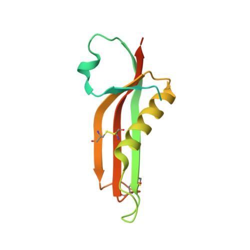
    The bacterial Rcs phosphorelay is a stress-induced defense mechanism that controls the expression of numerous genes, including those for capsular polysaccharides, motility, and virulence factors. It is a complex multicomponent system that includes the histidine kinase (RcsC) and the response regulator (RcsB) and also auxiliary proteins such as RcsF. RcsF is an outer membrane lipoprotein that transmits signals from the cell surface to RcsC. The physiological signals that activate RcsF and how RcsF interacts with RcsC remain unknown. Here, we report the three-dimensional structure of RcsF. The fold of the protein is characterized by the presence of a central 4-stranded β sheet, which is conserved in several other proteins, including the copper-binding domain of the amyloid precursor protein. RcsF, which contains four conserved cysteine residues, presents two nonconsecutive disulfides between Cys(74) and Cys(118) and between Cys(109) and Cys(124), respectively. These two disulfides are not functionally equivalent; the Cys(109)-Cys(124) disulfide is particularly important for the assembly of an active RcsF. Moreover, we show that formation of the nonconsecutive disulfides of RcsF depends on the periplasmic disulfide isomerase DsbC. We trapped RcsF in a mixed disulfide complex with DsbC, and we show that deletion of dsbC prevents the activation of the Rcs phosphorelay by signals that function through RcsF. The three-dimensional structure of RcsF provides the structural basis to understand how this protein triggers the Rcs signaling cascade.


  • Organizational Affiliation

    Welbio (Walloon Excellence in Life Sciences and Biotechnology), Université Catholique de Louvain, B-1200 Brussels, Belgium.


Macromolecules
Find similar proteins by:  (by identity cutoff)  |  3D Structure
Entity ID: 1
MoleculeChains Sequence LengthOrganismDetailsImage
PUTATIVE OUTER MEMBRANE PROTEIN, SIGNAL128Escherichia coli K-12Mutation(s): 0 
UniProt
Find proteins for P69411 (Escherichia coli (strain K12))
Explore P69411 
Go to UniProtKB:  P69411
Entity Groups  
Sequence Clusters30% Identity50% Identity70% Identity90% Identity95% Identity100% Identity
UniProt GroupP69411
Sequence Annotations
Expand
  • Reference Sequence
Small Molecules
Ligands 2 Unique
IDChains Name / Formula / InChI Key2D Diagram3D Interactions
I3C
Query on I3C

Download Ideal Coordinates CCD File 
B [auth A]5-amino-2,4,6-triiodobenzene-1,3-dicarboxylic acid
C8 H4 I3 N O4
JEZJSNULLBSYHV-UHFFFAOYSA-N
SO4
Query on SO4

Download Ideal Coordinates CCD File 
C [auth A]SULFATE ION
O4 S
QAOWNCQODCNURD-UHFFFAOYSA-L
Experimental Data & Validation

Experimental Data

  • Method: X-RAY DIFFRACTION
  • Resolution: 2.00 Å
  • R-Value Free: 0.258 
  • R-Value Work: 0.196 
  • R-Value Observed: 0.199 
  • Space Group: P 31 2 1
Unit Cell:
Length ( Å )Angle ( ˚ )
a = 55.714α = 90
b = 55.714β = 90
c = 61.679γ = 120
Software Package:
Software NamePurpose
REFMACrefinement
XDSdata reduction
XSCALEdata scaling
Auto-Rickshawphasing

Structure Validation

View Full Validation Report


Ligand Structure Quality Assessment 


Entry History 

Deposition Data

Revision History  (Full details and data files)

  • Version 1.0: 2011-03-16
    Type: Initial release
  • Version 1.1: 2011-05-12
    Changes: Version format compliance
  • Version 1.2: 2011-07-13
    Changes: Version format compliance
  • Version 1.3: 2019-05-08
    Changes: Data collection, Derived calculations, Experimental preparation, Other
  • Version 1.4: 2019-07-17
    Changes: Data collection